

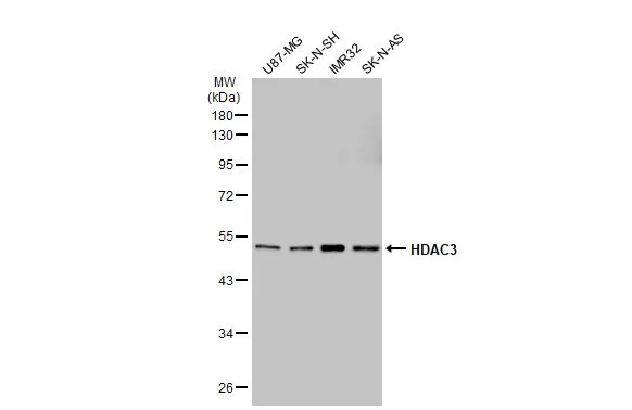
Various whole cell extracts (30 μg) were separated by 10% SDS-PAGE, and the membrane was blotted with HDAC3 antibody [GT1144] (GTX00834) diluted at 1:1000. The HRP-conjugated anti-rabbit IgG antibody (GTX213110-01) was used to detect the primary antibody, and the signal was developed with Trident ECL plus-Enhanced.
![WB analysis of various samples using GTX00834 HDAC3 antibody [GT1144]. Dilution : 1:1000 Loading : 25 μg](https://www.genetex.com/upload/website/prouct_img/normal/GTX00834/GTX00834_20200508_WB_w_23053121_289.webp "WB analysis of various samples using GTX00834 HDAC3 antibody [GT1144]. Dilution : 1:1000 Loading : 25 μg")
Various whole cell extracts (30 μg) were separated by 10% SDS-PAGE, and the membrane was blotted with HDAC3 antibody [GT1144] (GTX00834) diluted at 1:1000. The HRP-conjugated anti-rabbit IgG antibody (GTX213110-01) was used to detect the primary antibody, and the signal was developed with Trident ECL plus-Enhanced.
HDAC3 antibody [GT1144]
GTX00834
ApplicationsWestern Blot
Product group Antibodies
ReactivityHuman, Mouse, Rat
TargetHDAC3
Overview
- SupplierGeneTex
- Product NameHDAC3 antibody [GT1144]
- Delivery Days Customer9
- Application Supplier NoteWB: 1:500 - 1:2000. *Optimal dilutions/concentrations should be determined by the researcher.Not tested in other applications.
- ApplicationsWestern Blot
- CertificationResearch Use Only
- ClonalityMonoclonal
- Clone IDGT1144
- ConjugateUnconjugated
- Gene ID8841
- Target nameHDAC3
- Target descriptionhistone deacetylase 3
- Target synonymsHD3, KDAC3, RPD3, RPD3-2, histone deacetylase 3, SMAP45, protein deacetylase HDAC3, protein deacylase HDAC3
- HostRabbit
- IsotypeIgG
- Protein IDO15379
- Protein NameHistone deacetylase 3
- Scientific DescriptionHistones play a critical role in transcriptional regulation, cell cycle progression, and developmental events. Histone acetylation/deacetylation alters chromosome structure and affects transcription factor access to DNA. The protein encoded by this gene belongs to the histone deacetylase/acuc/apha family. It has histone deacetylase activity and represses transcription when tethered to a promoter. It may participate in the regulation of transcription through its binding with the zinc-finger transcription factor YY1. This protein can also down-regulate p53 function and thus modulate cell growth and apoptosis. This gene is regarded as a potential tumor suppressor gene. [provided by RefSeq, Jul 2008]
- ReactivityHuman, Mouse, Rat
- Storage Instruction-20°C or -80°C,2°C to 8°C
- UNSPSC41116161
Datasheet
Related products
Product group Antibodies
Anti-HDAC3 AntibodyA95226
ApplicationsImmunoFluorescence, Western Blot, ELISA, ImmunoHistoChemistry
ReactivityHuman, Mouse, Rat
- SizePrice

Product group Antibodies
Anti-HDAC3 Antibody Picoband(r)A00839-CARRIER-FREE
ApplicationsFlow Cytometry, Western Blot, ELISA
ReactivityHuman, Mouse, Rat
TargetHDAC3
- SizePrice
Product group Antibodies
Anti-HDAC3 Antibody144-02139
ApplicationsImmunoFluorescence, ImmunoPrecipitation, Western Blot, ChIP Chromatin ImmunoPrecipitation, ImmunoHistoChemistry
ReactivityHuman, Mouse, Rat
TargetHDAC3
- SizePrice
Product group Antibodies
ApplicationsImmunoFluorescence, ChIP Chromatin ImmunoPrecipitation
ReactivityHuman
TargetHDAC3
- SizePrice
Product group Antibodies
ApplicationsFlow Cytometry, Western Blot, ImmunoCytoChemistry
ReactivityHuman, Mouse, Rat
TargetHDAC3
- SizePrice
Product group Antibodies
HDAC3 AntibodyCSB-PA002880
ApplicationsImmunoFluorescence, Western Blot, ELISA, ImmunoHistoChemistry
ReactivityHuman, Mouse, Rat
TargetHDAC3
- SizePrice
Product group Antibodies

Goat anti-HDAC3EB12818
ApplicationsWestern Blot, ELISA
ReactivityBovine, Canine, Human, Mouse, Porcine, Rat
TargetHDAC3
- SizePrice
Product group Antibodies
HDAC3 Polyclonal AntibodyCAC13761
ApplicationsImmunoFluorescence, Western Blot, ChIP Chromatin ImmunoPrecipitation, ELISA, ImmunoHistoChemistry
ReactivityMouse
TargetHDAC3
- SizePrice
Product group Antibodies

HDAC3 Antibody (phospho-Ser424)LS-C359014
ApplicationsImmunoFluorescence, Western Blot, ImmunoCytoChemistry, ImmunoHistoChemistry, ImmunoHistoChemistry Paraffin
ReactivityChicken, Human, Mouse, Rat
TargetHDAC3
- SizePrice
![IHC-P analysis of human esophagus cancer (left) and breast carcinoma tissue (right) using GTX83172 HDAC3 antibody [3A7B5].](https://www.genetex.com/upload/website/prouct_img/normal/GTX83172/GTX83172_20170912_IHC-P_w_23061322_791.webp)
Product group Antibodies
HDAC3 antibody [3A7B5]GTX83172
ApplicationsWestern Blot, ELISA, ImmunoHistoChemistry, ImmunoHistoChemistry Paraffin
ReactivityHuman
TargetHDAC3
- SizePrice