

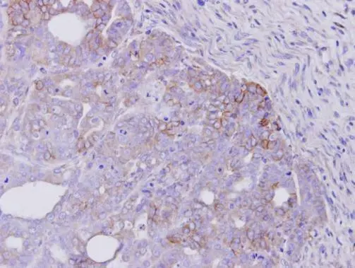
Immunohistochemical analysis of paraffin-embedded NCIN87 xenograft, using ERBB4(v-erb-A)(GTX111276) antibody at 1:500 dilution.
Antigen Retrieval: Trilogy? (EDTA based, pH 8.0) buffer, 15min
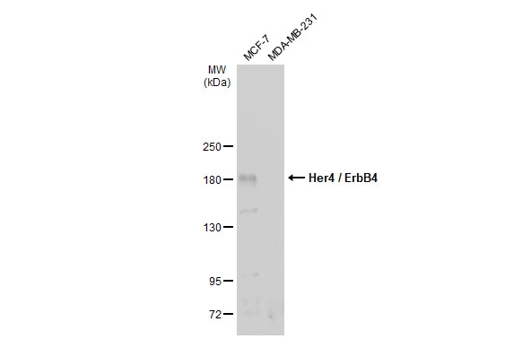
![Immunoprecipitation of Her4 / ErbB4 protein from MCF-7 whole cell extract using 4.5 μg of Her4 / ErbB4 antibody [C1C3] (GTX111276). Western blot analysis was performed using Her4 / ErbB4 antibody [C1C3] (GTX111276) and diluted at 1:500. EasyBlot HRP-conjugated anti rabbit IgG antibody (GTX221666-01) was used to detect the primary antibody.](https://www.genetex.com/upload/website/prouct_img/normal/GTX111276/GTX111276_40058_20170714_IP_w_23060500_975.webp "Immunoprecipitation of Her4 / ErbB4 protein from MCF-7 whole cell extract using 4.5 μg of Her4 / ErbB4 antibody [C1C3] (GTX111276). Western blot analysis was performed using Her4 / ErbB4 antibody [C1C3] (GTX111276) and diluted at 1:500. EasyBlot HRP-conjugated anti rabbit IgG antibody (GTX221666-01) was used to detect the primary antibody.")
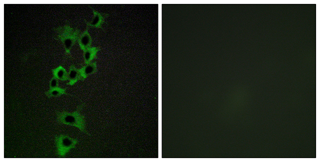
![ERBB4 antibody [C1C3] detects ERBB4 protein at cytoplasm and nucleus by immunofluorescent analysis. Sample: MCF-7 cells were fixed in ice-cold MeOH for 5 min. Green: ERBB4 protein stained by ERBB4 antibody [C1C3] (GTX111276) diluted at 1:500. Blue: Hoechst 33342 staining.](https://www.genetex.com/upload/website/prouct_img/normal/GTX111276/GTX111276_40058_IFA_w_23060500_659.webp "ERBB4 antibody [C1C3] detects ERBB4 protein at cytoplasm and nucleus by immunofluorescent analysis. Sample: MCF-7 cells were fixed in ice-cold MeOH for 5 min. Green: ERBB4 protein stained by ERBB4 antibody [C1C3] (GTX111276) diluted at 1:500. Blue: Hoechst 33342 staining.")
![Various tissue extracts (50 μg) were separated by 5% SDS-PAGE, and the membrane was blotted with Her4 / ErbB4 antibody [C1C3] (GTX111276) diluted at 1:1000. The HRP-conjugated anti-rabbit IgG antibody (GTX213110-01) was used to detect the primary antibody.](https://www.genetex.com/upload/website/prouct_img/normal/GTX111276/GTX111276_42907_20170721_WB_M_R_w_23060500_769.webp "Various tissue extracts (50 μg) were separated by 5% SDS-PAGE, and the membrane was blotted with Her4 / ErbB4 antibody [C1C3] (GTX111276) diluted at 1:1000. The HRP-conjugated anti-rabbit IgG antibody (GTX213110-01) was used to detect the primary antibody.")
![Non-transfected (–) and transfected (+) 293T whole cell extracts (30 μg) were separated by 5% SDS-PAGE, and the membrane was blotted with Her4 / ErbB4 antibody [C1C3] (GTX111276) diluted at 1:5000. The HRP-conjugated anti-rabbit IgG antibody (GTX213110-01) was used to detect the primary antibody.](https://www.genetex.com/upload/website/prouct_img/normal/GTX111276/GTX111276_42907_20180928_WB_B_w_23060500_931.webp "Non-transfected (–) and transfected (+) 293T whole cell extracts (30 μg) were separated by 5% SDS-PAGE, and the membrane was blotted with Her4 / ErbB4 antibody [C1C3] (GTX111276) diluted at 1:5000. The HRP-conjugated anti-rabbit IgG antibody (GTX213110-01) was used to detect the primary antibody.")
![Whole cell extract (30 μg) was separated by 5% SDS-PAGE, and the membrane was blotted with Her4 / ErbB4 antibody [C1C3] (GTX111276) diluted at 1:1000. The HRP-conjugated anti-rabbit IgG antibody (GTX213110-01) was used to detect the primary antibody.](https://www.genetex.com/upload/website/prouct_img/normal/GTX111276/GTX111276_43852_20200306_WB_w_23060500_484.webp "Whole cell extract (30 μg) was separated by 5% SDS-PAGE, and the membrane was blotted with Her4 / ErbB4 antibody [C1C3] (GTX111276) diluted at 1:1000. The HRP-conjugated anti-rabbit IgG antibody (GTX213110-01) was used to detect the primary antibody.")
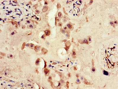
Immunohistochemical analysis of paraffin-embedded NCIN87 xenograft, using ERBB4(v-erb-A)(GTX111276) antibody at 1:500 dilution.
Antigen Retrieval: Trilogy? (EDTA based, pH 8.0) buffer, 15min
Her4 / ErbB4 antibody [C1C3]
GTX111276
ApplicationsImmunoFluorescence, ImmunoPrecipitation, Western Blot, ImmunoCytoChemistry, ImmunoHistoChemistry, ImmunoHistoChemistry Paraffin
Product group Antibodies
ReactivityHuman, Mouse, Rat
TargetERBB4
Overview
- SupplierGeneTex
- Product NameHer4 / ErbB4 antibody [C1C3]
- Delivery Days Customer9
- Application Supplier NoteWB: 1:1000-1:10000. ICC/IF: 1:100-1:1000. IHC-P: 1:100-1:1000. IP: 1:100-1:500. *Optimal dilutions/concentrations should be determined by the researcher.Not tested in other applications.
- ApplicationsImmunoFluorescence, ImmunoPrecipitation, Western Blot, ImmunoCytoChemistry, ImmunoHistoChemistry, ImmunoHistoChemistry Paraffin
- CertificationResearch Use Only
- ClonalityPolyclonal
- Concentration0.97 mg/ml
- ConjugateUnconjugated
- Gene ID2066
- Target nameERBB4
- Target descriptionerb-b2 receptor tyrosine kinase 4
- Target synonymsALS19, HER4, p180erbB4, receptor tyrosine-protein kinase erbB-4, avian erythroblastic leukemia viral (v-erb-b2) oncogene homolog 4, human epidermal growth factor receptor 4, proto-oncogene-like protein c-ErbB-4, tyrosine kinase-type cell surface receptor HER4, v-erb-a erythroblastic leukemia viral oncogene homolog 4, v-erb-b2 avian erythroblastic leukemia viral oncogene homolog 4
- HostRabbit
- IsotypeIgG
- Protein IDQ15303
- Protein NameReceptor tyrosine-protein kinase erbB-4
- Scientific DescriptionThis gene is a member of the Tyr protein kinase family and the epidermal growth factor receptor subfamily. It encodes a single-pass type I membrane protein with multiple cysteine rich domains, a transmembrane domain, a tyrosine kinase domain, a phosphotidylinositol-3 kinase binding site and a PDZ domain binding motif. The protein binds to and is activated by neuregulins and other factors and induces a variety of cellular responses including mitogenesis and differentiation. Multiple proteolytic events allow for the release of a cytoplasmic fragment and an extracellular fragment. Mutations in this gene have been associated with cancer. Alternatively spliced variants which encode different protein isoforms have been described; however, not all variants have been fully characterized. [provided by RefSeq]
- ReactivityHuman, Mouse, Rat
- Storage Instruction-20°C or -80°C,2°C to 8°C
- UNSPSC41116161
Datasheet
Related products
Product group Antibodies
Anti-HER4 AntibodyA96182
ApplicationsImmunoFluorescence, ELISA, ImmunoHistoChemistry
ReactivityHuman, Mouse, Rat
- SizePrice
Product group Antibodies

Anti-HER4 [B6 (HER4.B6)]Ab02661-10.0
ApplicationsELISA
ReactivityHuman
TargetERBB4
- SizePrice
Product group Antibodies
Anti-ERBB4 Antibody144-00749
ApplicationsWestern Blot
ReactivityHuman, Rat
TargetERBB4
- SizePrice

Product group Antibodies
Anti-ERBB4 Antibody Picoband(r)A00296-1-CARRIER-FREE
ApplicationsWestern Blot, ELISA, ImmunoHistoChemistry
ReactivityHuman, Mouse, Rat
TargetERBB4
- SizePrice
Product group Antibodies
ErbB4 Recombinant AntibodyBSM-61882R
ApplicationsFlow Cytometry, ImmunoFluorescence, ImmunoPrecipitation, Western Blot, ImmunoCytoChemistry
ReactivityHuman, Mouse, Rat
TargetERBB4
- SizePrice
Product group Antibodies
ERBB4 AntibodyCSB-PA007766LA01HU
ApplicationsImmunoFluorescence, ELISA, ImmunoHistoChemistry
ReactivityHuman
TargetERBB4
- SizePrice
Product group Antibodies
ApplicationsImmunoPrecipitation, Western Blot, ImmunoCytoChemistry, ImmunoHistoChemistry
ReactivityMouse, Rat
TargetERBB4
- SizePrice
Product group Antibodies
Her4 / ErbB4 antibodyGTX134351
ApplicationsWestern Blot
ReactivityHuman
TargetERBB4
- SizePrice
![IHC-P analysis of mouse placenta tissue using GTX80811 Her4 / ErbB4 antibody [HFR1]. Right : Primary antibody Left : Negative control without primary antibody Antigen retrieval : 10mM sodium citrate (pH 6.0), microwaved for 8-15 min Dilution : 1:100](https://www.genetex.com/upload/website/prouct_img/normal/GTX80811/GTX80811_1418_IHC-P_w_23061322_116.webp)
Product group Antibodies
References
Her4 / ErbB4 antibody [HFR1]GTX80811
ApplicationsFlow Cytometry, ImmunoFluorescence, ImmunoPrecipitation, Western Blot, ImmunoCytoChemistry, ImmunoHistoChemistry, ImmunoHistoChemistry Frozen, ImmunoHistoChemistry Paraffin
ReactivityHuman, Mouse
TargetERBB4
- SizePrice