

IHC-P analysis of mouse placenta tissue using GTX80811 Her4 / ErbB4 antibody [HFR1]. Right : Primary antibody Left : Negative control without primary antibody Antigen retrieval : 10mM sodium citrate (pH 6.0), microwaved for 8-15 min Dilution : 1:100
![IHC-P analysis of human heart tissue using GTX80811 Her4 / ErbB4 antibody [HFR1]. Left : Primary antibody Right : Negative control without primary antibody Antigen retrieval : heat induced antigen retrieval was performed using 10mM sodium citrate (pH6.0) buffer, microwaved for 8-15 minutes Dilution : 1:200](https://www.genetex.com/upload/website/prouct_img/normal/GTX80811/GTX80811_1414_IHC-P_w_23061322_183.webp "IHC-P analysis of human heart tissue using GTX80811 Her4 / ErbB4 antibody [HFR1]. Left : Primary antibody Right : Negative control without primary antibody Antigen retrieval : heat induced antigen retrieval was performed using 10mM sodium citrate (pH6.0) buffer, microwaved for 8-15 minutes Dilution : 1:200")
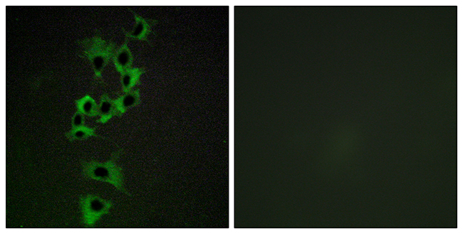
![ICC/IF analysis of A431 cells using GTX80811 Her4 / ErbB4 antibody [HFR1]. Cells were probed without (left) or with(right) an antibody. Green : Primary antibody Blue : Nuclei Red : Actin Fixation : Formalin Permeabilization : 0.1% Triton X-100 in TBS for 5-10 minute Dilution : 1:50 and incubated overnight](https://www.genetex.com/upload/website/prouct_img/normal/GTX80811/GTX80811_907_ICC-IF_w_23061322_634.webp "ICC/IF analysis of A431 cells using GTX80811 Her4 / ErbB4 antibody [HFR1]. Cells were probed without (left) or with(right) an antibody. Green : Primary antibody Blue : Nuclei Red : Actin Fixation : Formalin Permeabilization : 0.1% Triton X-100 in TBS for 5-10 minute Dilution : 1:50 and incubated overnight")
![ICC/IF analysis of SK-BR-3 cells using GTX80811 Her4 / ErbB4 antibody [HFR1]. Cells were probed without (left) or with(right) an antibody. Green : Primary antibody Blue : Nuclei Red : Actin Fixation : Formalin Permeabilization : 0.1% Triton X-100 in TBS for 5-10 minute Dilution : 1:50 and incubated overnight](https://www.genetex.com/upload/website/prouct_img/normal/GTX80811/GTX80811_909_ICC-IF_w_23061322_120.webp "ICC/IF analysis of SK-BR-3 cells using GTX80811 Her4 / ErbB4 antibody [HFR1]. Cells were probed without (left) or with(right) an antibody. Green : Primary antibody Blue : Nuclei Red : Actin Fixation : Formalin Permeabilization : 0.1% Triton X-100 in TBS for 5-10 minute Dilution : 1:50 and incubated overnight")
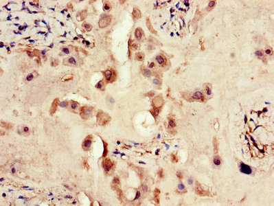
![IHC-P analysis of human pancreas tissue using GTX80811 Her4 / ErbB4 antibody [HFR1]. Left : Primary antibody Right : Negative control without primary antibody Antigen retrieval : heat induced antigen retrieval was performed using 10mM sodium citrate (pH6.0) buffer, microwaved for 8-15 minutes Dilution : 1:200](https://www.genetex.com/upload/website/prouct_img/normal/GTX80811/GTX80811_1415_IHC-P_w_23061322_981.webp "IHC-P analysis of human pancreas tissue using GTX80811 Her4 / ErbB4 antibody [HFR1]. Left : Primary antibody Right : Negative control without primary antibody Antigen retrieval : heat induced antigen retrieval was performed using 10mM sodium citrate (pH6.0) buffer, microwaved for 8-15 minutes Dilution : 1:200")
![FACS analysis of MCF-7 cells using GTX80811 Her4 / ErbB4 antibody [HFR1] compared to an isotype control (blue). Dilution : 1ug/test for 40 min at room temperature Fixation : 2% paraformaldehyde](https://www.genetex.com/upload/website/prouct_img/normal/GTX80811/GTX80811_221_FACS_w_23061322_139.webp "FACS analysis of MCF-7 cells using GTX80811 Her4 / ErbB4 antibody [HFR1] compared to an isotype control (blue). Dilution : 1ug/test for 40 min at room temperature Fixation : 2% paraformaldehyde")
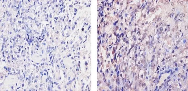
![IHC-P analysis of human breast carcinoma tissue using GTX80811 Her4 / ErbB4 antibody [HFR1]. Left : Primary antibody Right : Negative control without primary antibody Antigen retrieval : heat induced antigen retrieval was performed using 10mM sodium citrate (pH6.0) buffer, microwaved for 8-15 minutes Dilution : 1:20](https://www.genetex.com/upload/website/prouct_img/normal/GTX80811/GTX80811_1413_IHC-P_w_23061322_826.webp "IHC-P analysis of human breast carcinoma tissue using GTX80811 Her4 / ErbB4 antibody [HFR1]. Left : Primary antibody Right : Negative control without primary antibody Antigen retrieval : heat induced antigen retrieval was performed using 10mM sodium citrate (pH6.0) buffer, microwaved for 8-15 minutes Dilution : 1:20")
![FACS analysis of Hela cells using GTX80811 Her4 / ErbB4 antibody [HFR1] compared to an isotype control (blue). Dilution : 1ug/test for 40 min at room temperature Fixation : 2% paraformaldehyde](https://www.genetex.com/upload/website/prouct_img/normal/GTX80811/GTX80811_220_FACS_w_23061322_163.webp "FACS analysis of Hela cells using GTX80811 Her4 / ErbB4 antibody [HFR1] compared to an isotype control (blue). Dilution : 1ug/test for 40 min at room temperature Fixation : 2% paraformaldehyde")
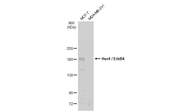
![WB analysis of 25 ug of MDA-MB-453 (Lane 1) and T47D (Lane 2) cell lysates using GTX80811 Her4 / ErbB4 antibody [HFR1]. Dilution : 1:300](https://www.genetex.com/upload/website/prouct_img/normal/GTX80811/GTX80811_1957_WB_w_23061322_561.webp "WB analysis of 25 ug of MDA-MB-453 (Lane 1) and T47D (Lane 2) cell lysates using GTX80811 Her4 / ErbB4 antibody [HFR1]. Dilution : 1:300")
![FACS analysis of NIH-3T3 cells using GTX80811 Her4 / ErbB4 antibody [HFR1] compared to an isotype control (blue). Dilution : 0.5ug/test for 40 min at room temperature Fixation : 2% paraformaldehyde](https://www.genetex.com/upload/website/prouct_img/normal/GTX80811/GTX80811_222_FACS_w_23061322_943.webp "FACS analysis of NIH-3T3 cells using GTX80811 Her4 / ErbB4 antibody [HFR1] compared to an isotype control (blue). Dilution : 0.5ug/test for 40 min at room temperature Fixation : 2% paraformaldehyde")
IHC-P analysis of mouse placenta tissue using GTX80811 Her4 / ErbB4 antibody [HFR1]. Right : Primary antibody Left : Negative control without primary antibody Antigen retrieval : 10mM sodium citrate (pH 6.0), microwaved for 8-15 min Dilution : 1:100
Her4 / ErbB4 antibody [HFR1]
GTX80811
ApplicationsFlow Cytometry, ImmunoFluorescence, ImmunoPrecipitation, Western Blot, ImmunoCytoChemistry, ImmunoHistoChemistry, ImmunoHistoChemistry Frozen, ImmunoHistoChemistry Paraffin
Product group Antibodies
ReactivityHuman, Mouse
TargetERBB4
Overview
- SupplierGeneTex
- Product NameHer4 / ErbB4 antibody [HFR1]
- Delivery Days Customer9
- Application Supplier NoteWB: 1:100-1:500. ICC/IF: 1:50. IHC-P: 1:100. FCM: 0.5-2 microg/test. *Optimal dilutions/concentrations should be determined by the researcher.Not tested in other applications.
- ApplicationsFlow Cytometry, ImmunoFluorescence, ImmunoPrecipitation, Western Blot, ImmunoCytoChemistry, ImmunoHistoChemistry, ImmunoHistoChemistry Frozen, ImmunoHistoChemistry Paraffin
- CertificationResearch Use Only
- ClonalityMonoclonal
- Clone IDHFR1
- Concentration1 mg/ml
- ConjugateUnconjugated
- Gene ID2066
- Target nameERBB4
- Target descriptionerb-b2 receptor tyrosine kinase 4
- Target synonymsALS19, HER4, p180erbB4, receptor tyrosine-protein kinase erbB-4, avian erythroblastic leukemia viral (v-erb-b2) oncogene homolog 4, human epidermal growth factor receptor 4, proto-oncogene-like protein c-ErbB-4, tyrosine kinase-type cell surface receptor HER4, v-erb-a erythroblastic leukemia viral oncogene homolog 4, v-erb-b2 avian erythroblastic leukemia viral oncogene homolog 4
- HostMouse
- IsotypeIgG2b
- Protein IDQ15303
- Protein NameReceptor tyrosine-protein kinase erbB-4
- Scientific DescriptionThis gene is a member of the Tyr protein kinase family and the epidermal growth factor receptor subfamily. It encodes a single-pass type I membrane protein with multiple cysteine rich domains, a transmembrane domain, a tyrosine kinase domain, a phosphotidylinositol-3 kinase binding site and a PDZ domain binding motif. The protein binds to and is activated by neuregulins and other factors and induces a variety of cellular responses including mitogenesis and differentiation. Multiple proteolytic events allow for the release of a cytoplasmic fragment and an extracellular fragment. Mutations in this gene have been associated with cancer. Alternatively spliced variants which encode different protein isoforms have been described; however, not all variants have been fully characterized. [provided by RefSeq, Jul 2008]
- ReactivityHuman, Mouse
- Storage Instruction-20°C or -80°C,2°C to 8°C
- UNSPSC41116161
References
- Dysregulation of ErbB4 Signaling Pathway in the Dorsal Hippocampus after Neonatal Hypoxia-Ischemia and Late Deficits in PV(+) Interneurons, Synaptic Plasticity and Working Memory.Read this paper
Datasheet
Related products
Product group Antibodies
Anti-HER4 AntibodyA96182
ApplicationsImmunoFluorescence, ELISA, ImmunoHistoChemistry
ReactivityHuman, Mouse, Rat
- SizePrice
Product group Antibodies

Anti-HER4 [B6 (HER4.B6)]Ab02661-10.0
ApplicationsELISA
ReactivityHuman
TargetERBB4
- SizePrice
Product group Antibodies
Anti-ERBB4 Antibody144-00749
ApplicationsWestern Blot
ReactivityHuman, Rat
TargetERBB4
- SizePrice

Product group Antibodies
Anti-ERBB4 Antibody Picoband(r)A00296-1-CARRIER-FREE
ApplicationsWestern Blot, ELISA, ImmunoHistoChemistry
ReactivityHuman, Mouse, Rat
TargetERBB4
- SizePrice
Product group Antibodies
ErbB4 Recombinant AntibodyBSM-61882R
ApplicationsFlow Cytometry, ImmunoFluorescence, ImmunoPrecipitation, Western Blot, ImmunoCytoChemistry
ReactivityHuman, Mouse, Rat
TargetERBB4
- SizePrice
Product group Antibodies
ERBB4 AntibodyCSB-PA007766LA01HU
ApplicationsImmunoFluorescence, ELISA, ImmunoHistoChemistry
ReactivityHuman
TargetERBB4
- SizePrice
Product group Antibodies
ApplicationsImmunoPrecipitation, Western Blot, ImmunoCytoChemistry, ImmunoHistoChemistry
ReactivityMouse, Rat
TargetERBB4
- SizePrice
Product group Antibodies
Her4 / ErbB4 antibodyGTX134351
ApplicationsWestern Blot
ReactivityHuman
TargetERBB4
- SizePrice

Product group Antibodies
Anti-ERBB4 AntibodyHPA012016
ApplicationsImmunoHistoChemistry
ReactivityHuman
TargetERBB4
- SizePrice