

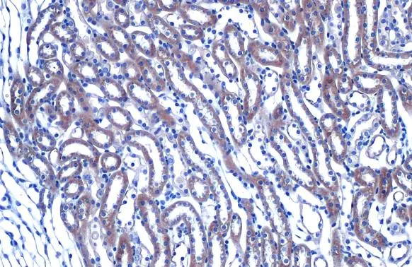
HHIP antibody [N3C2], Internal detects HHIP protein at cytoplasm by immunohistochemical analysis. Sample: Paraffin-embedded mouse kidney. HHIP stained by HHIP antibody [N3C2], Internal (GTX112584) diluted at 1:500. Antigen Retrieval: Citrate buffer, pH 6.0, 15 min
![HHIP antibody [N3C2], Internal antibody detects HHIP protein by immunofluorescent analysis. Sample: A431 cells were fixed in 4% paraformaldehyde for 15 min. Green: HHIP protein stained by HHIP antibody [N3C2], Internal antibody (GTX112584) diluted at 1:500. Blue: Hoechst 33342 staining. Scale bar = 10 μm.](https://www.genetex.com/upload/website/prouct_img/normal/GTX112584/GTX112584_40128_IFA_w_23060500_556.webp "HHIP antibody [N3C2], Internal antibody detects HHIP protein by immunofluorescent analysis. Sample: A431 cells were fixed in 4% paraformaldehyde for 15 min. Green: HHIP protein stained by HHIP antibody [N3C2], Internal antibody (GTX112584) diluted at 1:500. Blue: Hoechst 33342 staining. Scale bar = 10 μm.")
![HHIP antibody [N3C2], Internal detects HHIP protein at cytosol on human hepatoma by immunohistochemical analysis. Sample: Paraffin-embedded hepatoma. HHIP antibody [N3C2], Internal (GTX112584) dilution: 1:500.
Antigen Retrieval: Trilogy? (EDTA based, pH 8.0) buffer, 15min](https://www.genetex.com/upload/website/prouct_img/normal/GTX112584/GTX112584_40128_IHC_w_23060500_355.webp "HHIP antibody [N3C2], Internal detects HHIP protein at cytosol on human hepatoma by immunohistochemical analysis. Sample: Paraffin-embedded hepatoma. HHIP antibody [N3C2], Internal (GTX112584) dilution: 1:500.
Antigen Retrieval: Trilogy? (EDTA based, pH 8.0) buffer, 15min")
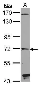
![Non-transfected (–) and transfected (+) 293T whole cell extracts (30 μg) were separated by 7.5% SDS-PAGE, and the membrane was blotted with HHIP antibody [N3C2], Internal (GTX112584) diluted at 1:5000. The HRP-conjugated anti-rabbit IgG antibody (GTX213110-01) was used to detect the primary antibody.](https://www.genetex.com/upload/website/prouct_img/normal/GTX112584/GTX112584_41787_20200306_WB_B_w_23060500_129.webp "Non-transfected (–) and transfected (+) 293T whole cell extracts (30 μg) were separated by 7.5% SDS-PAGE, and the membrane was blotted with HHIP antibody [N3C2], Internal (GTX112584) diluted at 1:5000. The HRP-conjugated anti-rabbit IgG antibody (GTX213110-01) was used to detect the primary antibody.")
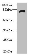
![Whole cell extract (30 μg) was separated by 7.5% SDS-PAGE, and the membrane was blotted with HHIP antibody [N3C2], Internal (GTX112584) diluted at 1:1000. The HRP-conjugated anti-rabbit IgG antibody (GTX213110-01) was used to detect the primary antibody.](https://www.genetex.com/upload/website/prouct_img/normal/GTX112584/GTX112584_44741_20220715_WB_24043002_550.webp "Whole cell extract (30 μg) was separated by 7.5% SDS-PAGE, and the membrane was blotted with HHIP antibody [N3C2], Internal (GTX112584) diluted at 1:1000. The HRP-conjugated anti-rabbit IgG antibody (GTX213110-01) was used to detect the primary antibody.")
![Various whole cell extracts (30 μg) were separated by 7.5% SDS-PAGE, and the membrane was blotted with HHIP antibody [N3C2], Internal (GTX112584) diluted at 1:1000. The HRP-conjugated anti-rabbit IgG antibody (GTX213110-01) was used to detect the primary antibody. Corresponding RNA expression data for the same cell lines are based on Human Protein Atlas program.](https://www.genetex.com/upload/website/prouct_img/normal/GTX112584/GTX112584_44741_20220715_WB_TPM_watermark_24043002_532.webp "Various whole cell extracts (30 μg) were separated by 7.5% SDS-PAGE, and the membrane was blotted with HHIP antibody [N3C2], Internal (GTX112584) diluted at 1:1000. The HRP-conjugated anti-rabbit IgG antibody (GTX213110-01) was used to detect the primary antibody. Corresponding RNA expression data for the same cell lines are based on Human Protein Atlas program.")
HHIP antibody [N3C2], Internal detects HHIP protein at cytoplasm by immunohistochemical analysis. Sample: Paraffin-embedded mouse kidney. HHIP stained by HHIP antibody [N3C2], Internal (GTX112584) diluted at 1:500. Antigen Retrieval: Citrate buffer, pH 6.0, 15 min
HHIP antibody [N3C2], Internal
GTX112584
ApplicationsImmunoFluorescence, Western Blot, ImmunoCytoChemistry, ImmunoHistoChemistry, ImmunoHistoChemistry Paraffin
Product group Antibodies
ReactivityChicken, Human, Mouse
TargetHHIP
Overview
- SupplierGeneTex
- Product NameHHIP antibody [N3C2], Internal
- Delivery Days Customer9
- Application Supplier NoteWB: 1:500-1:3000. ICC/IF: 1:100-1:1000. IHC-P: 1:100-1:1000. *Optimal dilutions/concentrations should be determined by the researcher.Not tested in other applications.
- ApplicationsImmunoFluorescence, Western Blot, ImmunoCytoChemistry, ImmunoHistoChemistry, ImmunoHistoChemistry Paraffin
- CertificationResearch Use Only
- ClonalityPolyclonal
- Concentration1.33 mg/ml
- ConjugateUnconjugated
- Gene ID64399
- Target nameHHIP
- Target descriptionhedgehog interacting protein
- Target synonymsHIP, hedgehog-interacting protein
- HostRabbit
- IsotypeIgG
- Protein IDQ96QV1
- Protein NameHedgehog-interacting protein
- Scientific DescriptionThis gene encodes a protein similar to the mouse hedgehog-interacting protein, a regulatory component of the hedgehog signalling pathway. Members of the hedgehog family are evolutionarily conserved proteins which are involved in many fundamental processes in embryonic development, including anteroposterior patterns of limbs and regulation of left-right asymmetry. [provided by RefSeq]
- ReactivityChicken, Human, Mouse
- Storage Instruction-20°C or -80°C,2°C to 8°C
- UNSPSC12352203
References
- Kosla J, Dvorak M, Cermak V. Molecular analysis of the TGF-beta controlled gene expression program in chicken embryo dermal myofibroblasts. Gene. 2013,513(1):90-100. doi: 10.1016/j.gene.2012.10.069Read this paper
Datasheet
Related products
Product group Antibodies
Anti-HHIP (internal) Antibody107-11034
ApplicationsImmunoFluorescence, Western Blot, ImmunoCytoChemistry, ImmunoHistoChemistry, ImmunoHistoChemistry Paraffin
ReactivityChicken, Human, Mouse
TargetHHIP
- SizePrice
Product group Antibodies
Hhip Polyclonal AntibodyCAC10879
ApplicationsWestern Blot, ELISA, ImmunoHistoChemistry
ReactivityRat
TargetHHIP
- SizePrice
Product group Antibodies
HHIP Polyclonal AntibodyBS-12316R
ApplicationsImmunoFluorescence, Western Blot, ELISA, ImmunoCytoChemistry, ImmunoHistoChemistry, ImmunoHistoChemistry Frozen, ImmunoHistoChemistry Paraffin
ReactivityBovine, Canine, Chicken, Equine, Human, Mouse, Rat, Sheep
TargetHHIP
- SizePrice
Product group Antibodies

HHIP / HIP AntibodyLS-C812990
ApplicationsImmunoFluorescence, Western Blot, ImmunoHistoChemistry
ReactivityBovine, Canine, Human, Porcine
TargetHHIP
- SizePrice
Product group Antibodies
HHIP AntibodyCSB-PA850419ESR2HU
ApplicationsWestern Blot, ELISA, ImmunoHistoChemistry
ReactivityHuman, Rat
TargetHHIP
- SizePrice
Product group Antibodies
HHIP antibody [C1C3]GTX112565
ApplicationsWestern Blot
ReactivityHuman
TargetHHIP
- SizePrice
![HHIP antibody [HL1737] detects HHIP protein at cell membrane and cytoplasm by immunohistochemical analysis. Sample: Paraffin-embedded mouse intestine. HHIP stained by HHIP antibody [HL1737] (GTX637385) diluted at 1:100. Antigen Retrieval: Citrate buffer, pH 6.0, 15 min](https://www.genetex.com/upload/website/prouct_img/normal/GTX637385/GTX637385_T-44781_20220902_IHC-P_M_22101319_911.webp)
Product group Antibodies
HHIP antibody [HL1737]GTX637385
ApplicationsWestern Blot, ImmunoHistoChemistry, ImmunoHistoChemistry Paraffin
ReactivityHuman, Mouse
TargetHHIP
- SizePrice
![HHIP antibody [HL1969] detects HHIP protein at cell membrane and secreted by immunohistochemical analysis. Sample: Paraffin-embedded mouse kidney. HHIP stained by HHIP antibody [HL1969] (GTX637862) diluted at 1:100. Antigen Retrieval: Citrate buffer, pH 6.0, 15 min](https://www.genetex.com/upload/website/prouct_img/normal/GTX637862/GTX637862_T-44858_20221111_IHC-P_M_1_22122018_387.webp)
Product group Antibodies
HHIP antibody [HL1969]GTX637862
ApplicationsWestern Blot, ImmunoHistoChemistry, ImmunoHistoChemistry Paraffin
ReactivityHuman, Mouse
TargetHHIP
- SizePrice