

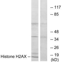
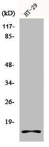
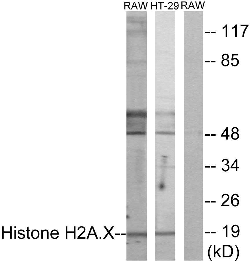
Western blot analysis of extracts from HT-29 cells, using Histone H2AX antibody.

Western blot analysis of extracts from HT-29 cells, using Histone H2AX antibody.
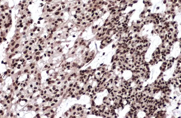
Histone H2AX Antibody
CSB-PA833019
ApplicationsImmunoFluorescence, Western Blot, ELISA
Product group Antibodies
ReactivityHuman
TargetH2AX
Overview
- SupplierCusabio
- Product NameHistone H2AX Antibody
- Delivery Days Customer20
- ApplicationsImmunoFluorescence, Western Blot, ELISA
- CertificationResearch Use Only
- ClonalityPolyclonal
- ConjugateUnconjugated
- Gene ID3014
- Target nameH2AX
- Target descriptionH2A.X variant histone
- Target synonymsH2A.X, H2A/X, H2AFX, histone H2AX, H2A histone family member X, H2AX histone, histone H2A.x
- HostRabbit
- IsotypeIgG
- Protein IDP16104
- Protein NameHistone H2AX
- Scientific DescriptionVariant histone H2A which replaces conventional H2A in a subset of nucleosomes. Nucleosomes wrap and compact DNA into chromatin, limiting DNA accessibility to the cellular machineries which require DNA as a template. Histones thereby play a central role in transcription regulation, DNA repair, DNA replication and chromosomal stability. DNA accessibility is regulated via a complex set of post-translational modifications of histones, also called histone code, and nucleosome remodeling. Required for checkpoint-mediated arrest of cell cycle progression in response to low doses of ionizing radiation and for efficient repair of DNA double strand breaks (DSBs) specifically when modified by C-terminal phosphorylation. The MGC Project Team; Genome Res. 14:2121-2127(2004). Rogakou E.P., J. Biol. Chem. 273:5858-5868(1998). Rogakou E.P., J. Biol. Chem. 275:9390-9395(2000).
- ReactivityHuman
- Storage Instruction-20°C or -80°C
- UNSPSC41116161
Related products
Product group Antibodies
Histone H2A.X AntibodyCSB-PA002920
ApplicationsImmunoFluorescence, Western Blot, ELISA, ImmunoHistoChemistry
ReactivityHuman
TargetH2AX
- SizePrice
Product group Antibodies
H2Afx Polyclonal AntibodyCAC07288
ApplicationsELISA, ImmunoHistoChemistry
TargetH2AX
- SizePrice

Product group Antibodies
Anti-Histone H2A.X/H2AFX Antibody Picoband(r)A00241-1-CARRIER-FREE
ApplicationsFlow Cytometry, ImmunoPrecipitation, Western Blot, ELISA, ImmunoHistoChemistry
ReactivityHuman, Mouse, Rat
TargetH2AX
- SizePrice
Product group Antibodies
Anti-Histone H2AX Antibody144-65468
ApplicationsImmunoFluorescence, Western Blot, ImmunoHistoChemistry
ReactivityHuman, Mouse, Rat
TargetH2AX
- SizePrice
Product group Antibodies
ApplicationsWestern Blot, ELISA, ImmunoHistoChemistry
ReactivityHuman, Mouse, Rat
- SizePrice

Product group Antibodies
Anti-H2AFX AntibodyAMAB91346
ApplicationsImmunoCytoChemistry, ImmunoHistoChemistry
ReactivityHuman
TargetH2AX
- SizePrice
Product group Antibodies

H2AFX / H2AX Antibody (C-Terminus)LS-C358767
ApplicationsImmunoFluorescence, Western Blot, ImmunoCytoChemistry, ImmunoHistoChemistry, ImmunoHistoChemistry Paraffin
ReactivityHuman, Mouse
TargetH2AX
- SizePrice
Product group Antibodies
References
ApplicationsFlow Cytometry, ImmunoFluorescence, Western Blot, ELISA, ImmunoCytoChemistry, ImmunoHistoChemistry, ImmunoHistoChemistry Frozen, ImmunoHistoChemistry Paraffin
ReactivityBovine, Canine, Equine, Human, Mouse, Porcine, Rabbit, Rat
TargetH2AX
- SizePrice
Product group Antibodies
Histone H2A.X antibodyGTX108272
ApplicationsImmunoFluorescence, ImmunoPrecipitation, Western Blot, ImmunoCytoChemistry, ImmunoHistoChemistry, ImmunoHistoChemistry Paraffin
ReactivityHuman, Mouse, Rat
TargetH2AX
- SizePrice