

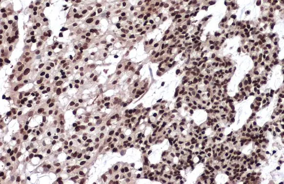
Histone H2A.X antibody detects Histone H2A.X protein at nucleus by immunohistochemical analysis. Sample: Paraffin-embedded human breast carcinoma. Histone H2A.X stained by Histone H2A.X antibody (GTX108272) diluted at 1:500. Antigen Retrieval: Citrate buffer, pH 6.0, 15 min
![Histone H2A.X antibody immunoprecipitates H2AFX protein in IP experiments. IP samples: Jurkat whole cell extract A. 40 μg Jurkat whole cell extract B. Control with 4 μg of preimmune Rabbit IgG C. Immunoprecipitation of H2AFX protein by 4 μg Histone H2A.X antibody (GTX108272) 15 % SDS-PAGE The immunoprecipitated H2AFX protein was detected by Histone H2A.X antibody (GTX108272) diluted at 1:2000. [EasyBlot anti-rabbit IgG (GTX221666-01) was used as a secondary reagent]](https://www.genetex.com/upload/website/prouct_img/normal/GTX108272/GTX108272_39778_IP_w_23060120_531.webp "Histone H2A.X antibody immunoprecipitates H2AFX protein in IP experiments. IP samples: Jurkat whole cell extract A. 40 μg Jurkat whole cell extract B. Control with 4 μg of preimmune Rabbit IgG C. Immunoprecipitation of H2AFX protein by 4 μg Histone H2A.X antibody (GTX108272) 15 % SDS-PAGE The immunoprecipitated H2AFX protein was detected by Histone H2A.X antibody (GTX108272) diluted at 1:2000. [EasyBlot anti-rabbit IgG (GTX221666-01) was used as a secondary reagent]")
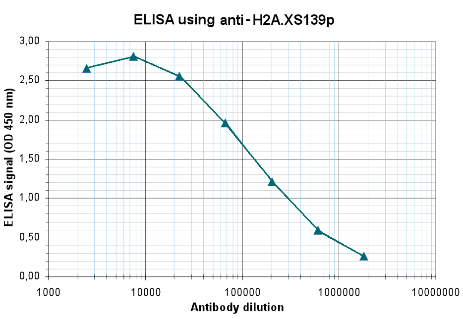
 dilution: 1:1000 The HRP-conjugated anti-rabbit IgG antibody (GTX213110-01) was used to detect the primary antibody.")
 diluted at 1:500.
Antigen Retrieval: Citrate buffer, pH 6.0, 15 min")
 diluted at 1:500.
Antigen Retrieval: Citrate buffer, pH 6.0, 15 min")
 diluted at 1:2000. Red: phalloidin, a cytoskeleton marker, diluted at 1:200. Scale bar= 10 μm.")
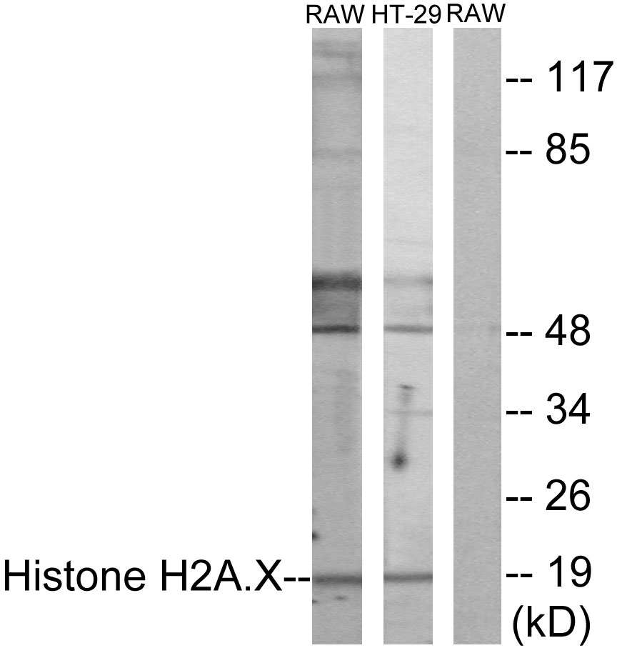
 and treated (+) HCT116 whole cell extracts (30 μg) were separated by 15% SDS-PAGE, and the membrane was blotted with Histone H2A.X antibody (GTX108272) diluted at 1:10000. The HRP-conjugated anti-rabbit IgG antibody (GTX213110-01) was used to detect the primary antibody.")
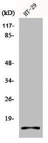
 dilution: 1:1000 The HRP-conjugated anti-rabbit IgG antibody (GTX213110-01) was used to detect the primary antibody.")
 dilution: 1:500.
Antigen Retrieval: Trilogy? (EDTA based, pH 8.0) buffer, 15min")
 of paraformaldehyde-fixed U2OS, using Histone H2A.X (GTX108272) antibody (Green) at 1:500 dilution. Alpha-tubulin filaments were labeled with GTX11304 (Red) at 1:2000.")
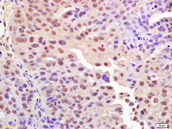
Histone H2A.X antibody detects Histone H2A.X protein at nucleus by immunohistochemical analysis. Sample: Paraffin-embedded human breast carcinoma. Histone H2A.X stained by Histone H2A.X antibody (GTX108272) diluted at 1:500. Antigen Retrieval: Citrate buffer, pH 6.0, 15 min
Histone H2A.X antibody
GTX108272
ApplicationsImmunoFluorescence, ImmunoPrecipitation, Western Blot, ImmunoCytoChemistry, ImmunoHistoChemistry, ImmunoHistoChemistry Paraffin
Product group Antibodies
ReactivityHuman, Mouse, Rat
TargetH2AX
Overview
- SupplierGeneTex
- Product NameHistone H2A.X antibody
- Delivery Days Customer9
- Application Supplier NoteWB: 1:500-1:20000. ICC/IF: 1:100-1:1000. IHC-P: 1:100-1:1000. IP: 1:100-1:500. *Optimal dilutions/concentrations should be determined by the researcher.Not tested in other applications.
- ApplicationsImmunoFluorescence, ImmunoPrecipitation, Western Blot, ImmunoCytoChemistry, ImmunoHistoChemistry, ImmunoHistoChemistry Paraffin
- CertificationResearch Use Only
- ClonalityPolyclonal
- Concentration0.19 mg/ml
- ConjugateUnconjugated
- Gene ID3014
- Target nameH2AX
- Target descriptionH2A.X variant histone
- Target synonymsH2A.X, H2A/X, H2AFX, histone H2AX, H2A histone family member X, H2AX histone, histone H2A.x
- HostRabbit
- IsotypeIgG
- Protein IDP16104
- Protein NameHistone H2AX
- Scientific DescriptionHistones are basic nuclear proteins that are responsible for the nucleosome structure of the chromosomal fiber in eukaryotes. Two molecules of each of the four core histones (H2A, H2B, H3, and H4) form an octamer, around which approximately 146 bp of DNA is wrapped in repeating units, called nucleosomes. The linker histone, H1, interacts with linker DNA between nucleosomes and functions in the compaction of chromatin into higher order structures. This gene encodes a member of the histone H2A family, and generates two transcripts through the use of the conserved stem-loop termination motif, and the polyA addition motif. [provided by RefSeq]
- ReactivityHuman, Mouse, Rat
- Storage Instruction-20°C or -80°C,2°C to 8°C
- UNSPSC41116161
Datasheet
Related products
Product group Antibodies
ApplicationsWestern Blot, ELISA, ImmunoHistoChemistry
ReactivityHuman, Mouse, Rat
- SizePrice
Product group Antibodies
ApplicationsDot Blot, Western Blot, ELISA
ReactivityHuman
TargetH2AX
- SizePrice
Product group Antibodies
Anti-Histone H2AX Antibody144-65468
ApplicationsImmunoFluorescence, Western Blot, ImmunoHistoChemistry
ReactivityHuman, Mouse, Rat
TargetH2AX
- SizePrice

Product group Antibodies
Anti-H2AFX AntibodyAMAB91346
ApplicationsImmunoCytoChemistry, ImmunoHistoChemistry
ReactivityHuman
TargetH2AX
- SizePrice

Product group Antibodies
Anti-Histone H2A.X/H2AFX Antibody Picoband(r)A00241-1-CARRIER-FREE
ApplicationsFlow Cytometry, ImmunoPrecipitation, Western Blot, ELISA, ImmunoHistoChemistry
ReactivityHuman, Mouse, Rat
TargetH2AX
- SizePrice
Product group Antibodies
References
ApplicationsFlow Cytometry, ImmunoFluorescence, Western Blot, ELISA, ImmunoCytoChemistry, ImmunoHistoChemistry, ImmunoHistoChemistry Frozen, ImmunoHistoChemistry Paraffin
ReactivityBovine, Canine, Equine, Human, Mouse, Porcine, Rabbit, Rat
TargetH2AX
- SizePrice
Product group Antibodies
Histone H2A.X AntibodyCSB-PA002920
ApplicationsImmunoFluorescence, Western Blot, ELISA, ImmunoHistoChemistry
ReactivityHuman
TargetH2AX
- SizePrice
Product group Antibodies
H2Afx Polyclonal AntibodyCAC07288
ApplicationsELISA, ImmunoHistoChemistry
TargetH2AX
- SizePrice
Product group Antibodies

H2AFX / H2AX Antibody (C-Terminus)LS-C358767
ApplicationsImmunoFluorescence, Western Blot, ImmunoCytoChemistry, ImmunoHistoChemistry, ImmunoHistoChemistry Paraffin
ReactivityHuman, Mouse
TargetH2AX
- SizePrice
![ICC/IF analysis of cells using GTX80694 Histone H2A.XS139ph (phospho Ser139) antibody [3F2].](https://www.genetex.com/upload/website/prouct_img/normal/GTX80694/GTX80694_2034_ICC-IF_w_23061322_855.webp)
Product group Antibodies
References
ApplicationsFlow Cytometry, ImmunoFluorescence, Western Blot, ELISA, ImmunoCytoChemistry, ImmunoHistoChemistry, ImmunoHistoChemistry Paraffin
ReactivityBovine, Human, Mouse
TargetH2AX
- SizePrice