

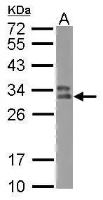
Western blot All lanes: HLA-DPB1 antibody at 2microg/ml Lane 1: sw1990 whole cell lysate Lane 2: HGC27 whole cell lysate Secondary Goat polyclonal to rabbit IgG at 1/10000 dilution Predicted band size: 30 kDa Observed band size: 30 kDa

Western blot All lanes: HLA-DPB1 antibody at 2microg/ml Lane 1: sw1990 whole cell lysate Lane 2: HGC27 whole cell lysate Secondary Goat polyclonal to rabbit IgG at 1/10000 dilution Predicted band size: 30 kDa Observed band size: 30 kDa
HLA-DPB1 Antibody
CSB-PA14809A0RB
ApplicationsWestern Blot, ELISA, ImmunoHistoChemistry
Product group Antibodies
ReactivityHuman
TargetHLA-DPB1
Overview
- SupplierCusabio
- Product NameHLA-DPB1 Antibody
- Delivery Days Customer20
- ApplicationsWestern Blot, ELISA, ImmunoHistoChemistry
- CertificationResearch Use Only
- ClonalityPolyclonal
- ConjugateUnconjugated
- Gene ID3115
- Target nameHLA-DPB1
- Target descriptionmajor histocompatibility complex, class II, DP beta 1
- Target synonymsDPB1, HLA-DP, HLA-DP1B, HLA-DPB, HLA class II histocompatibility antigen, DP beta 1 chain, HLA class II histocompatibility antigen, DP(W4) beta chain, HLA-DP histocompatibility type, beta-1 subunit, MHC HLA DPB1, MHC class II HLA-DP-beta-1, MHC class II antigen DPB1
- HostRabbit
- IsotypeIgG
- Protein IDP04440
- Protein NameHLA class II histocompatibility antigen, DP beta 1 chain
- Scientific DescriptionBinds peptides derived from antigens that access the endocytic route of antigen presenting cells (APC) and presents them on the cell surface for recognition by the CD4 T-cells. The peptide binding cleft accommodates peptides of 10-30 residues. The peptides presented by MHC class II molecules are generated mostly by degradation of proteins that access the endocytic route, where they are processed by lysosomal proteases and other hydrolases. Exogenous antigens that have been endocytosed by the APC are thus readily available for presentation via MHC II molecules, and for this reason this antigen presentation pathway is usually referred to as exogenous. As membrane proteins on their way to degradation in lysosomes as part of their normal turn-over are also contained in the endosomal/lysosomal compartments, exogenous antigens must compete with those derived from endogenous components. Autophagy is also a source of endogenous peptides, autophagosomes constitutively fuse with MHC class II loading compartments. In addition to APCs, other cells of the gastrointestinal tract, such as epithelial cells, express MHC class II molecules and CD74 and act as APCs, which is an unusual trait of the GI tract. To produce a MHC class II molecule that presents an antigen, three MHC class II molecules (heterodimers of an alpha and a beta chain) associate with a CD74 trimer in the ER to form a heterononamer. Soon after the entry of this complex into the endosomal/lysosomal system where antigen processing occurs, CD74 undergoes a sequential degradation by various proteases, including CTSS and CTSL, leaving a small fragment termed CLIP (class-II-associated invariant chain peptide). The removal of CLIP is facilitated by HLA-DM via direct binding to the alpha-beta-CLIP complex so that CLIP is released. HLA-DM stabilizes MHC class II molecules until primary high affinity antigenic peptides are bound. The MHC II molecule bound to a peptide is then transported to the cell membrane surface. In B-cells, the interaction between HLA-DM and MHC class II molecules is regulated by HLA-DO. Primary dendritic cells (DCs) also to express HLA-DO. Lysosomal miroenvironment has been implicated in the regulation of antigen loading into MHC II molecules, increased acidification produces increased proteolysis and efficient peptide loading.
- ReactivityHuman
- Storage Instruction-20°C or -80°C
- UNSPSC41116161
Related products
Product group Antibodies
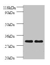
Anti-HLA-DPB1 AntibodyA30210
ApplicationsWestern Blot, ImmunoHistoChemistry
ReactivityHuman, Mouse, Rat
- SizePrice
Product group Antibodies
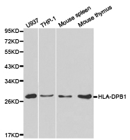
Anti-HLA-DPB1 Antibody144-01412
ApplicationsWestern Blot, ImmunoHistoChemistry
ReactivityHuman, Mouse, Rat
TargetHLA-DPB1
- SizePrice
Product group Antibodies

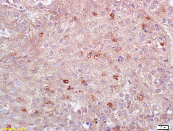
HLA-DPB1 AntibodyLS-C830586
ApplicationsELISA, ImmunoHistoChemistry
ReactivityHuman
TargetHLA-DPB1
- SizePrice

Product group Antibodies
Anti-HLA-DPB1 Antibody Picoband(r)A00487-CARRIER-FREE
ApplicationsWestern Blot, ImmunoHistoChemistry
ReactivityHuman
TargetHLA-DPB1
- SizePrice
Product group Antibodies
References
HLA-DPB1 Polyclonal AntibodyBS-4107R
ApplicationsFlow Cytometry, ImmunoFluorescence, Western Blot, ELISA, ImmunoCytoChemistry, ImmunoHistoChemistry, ImmunoHistoChemistry Frozen, ImmunoHistoChemistry Paraffin
ReactivityHuman, Mouse
TargetHLA-DPB1
- SizePrice
Product group Antibodies
HLA-DPB1 Polyclonal AntibodyCAC14140
ApplicationsWestern Blot, ELISA, ImmunoHistoChemistry
TargetHLA-DPB1
- SizePrice
Product group Antibodies
HLA-DPB1 antibodyGTX102763
ApplicationsWestern Blot
ReactivityHuman
TargetHLA-DPB1
- SizePrice

Product group Antibodies
Anti-HLA-DPB1 AntibodyHPA011078
ApplicationsWestern Blot, ImmunoHistoChemistry
ReactivityHuman
TargetHLA-DPB1
- SizePrice