

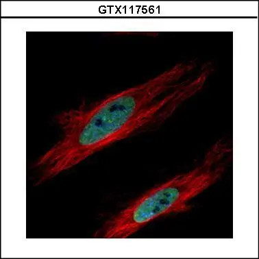
Confocal immunofluorescence analysis (Olympus FV10i) of paraformaldehyde-fixed HeLa, using HP1 gamma(GTX117561) antibody (Green) at 1:500 dilution. Alpha-tubulin filaments were labeled with GTX11304 (Red) at 1:2000.
 diluted at 1:500.
Antigen Retrieval: Citrate buffer, pH 6.0, 15 min")
 and HP1 gamma knockout (KO) HeLa cell extracts (30 μg) were separated by 15% SDS-PAGE, and the membrane was blotted with HP1 gamma antibody (GTX117561) diluted at 1:20000. The HRP-conjugated anti-rabbit IgG antibody (GTX213110-01) was used to detect the primary antibody.")
 15% SDS-PAGE The immunoprecipitated HP1 gamma protein was detected by HP1 gamma antibody (GTX117561) diluted at 1:1000. EasyBlot anti-rabbit IgG (GTX221666-01) was used as a secondary reagent.")
 diluted at 1:500.
Antigen Retrieval: Citrate buffer, pH 6.0, 15 min")
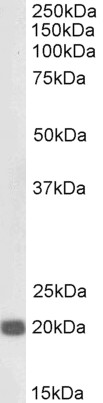
 A: HepG2 12% SDS PAGE GTX117561 diluted at 1:10000")
 antibody at 1:250 dilution.
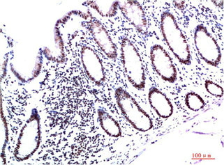
Antigen Retrieval: Citrate buffer, pH 6.0, 15 min")
Confocal immunofluorescence analysis (Olympus FV10i) of paraformaldehyde-fixed HeLa, using HP1 gamma(GTX117561) antibody (Green) at 1:500 dilution. Alpha-tubulin filaments were labeled with GTX11304 (Red) at 1:2000.
HP1 gamma antibody
GTX117561
ApplicationsImmunoFluorescence, ImmunoPrecipitation, Western Blot, ImmunoCytoChemistry, ImmunoHistoChemistry, ImmunoHistoChemistry Paraffin
Product group Antibodies
ReactivityHuman, Mouse
TargetCBX3
Overview
- SupplierGeneTex
- Product NameHP1 gamma antibody
- Delivery Days Customer9
- Application Supplier NoteWB: 1:5000-1:20000. ICC/IF: 1:100-1:1000. IHC-P: 1:100-1:1000. IP: 1:100-1:500. *Optimal dilutions/concentrations should be determined by the researcher.Not tested in other applications.
- ApplicationsImmunoFluorescence, ImmunoPrecipitation, Western Blot, ImmunoCytoChemistry, ImmunoHistoChemistry, ImmunoHistoChemistry Paraffin
- CertificationResearch Use Only
- ClonalityPolyclonal
- Concentration1 mg/ml
- ConjugateUnconjugated
- Gene ID11335
- Target nameCBX3
- Target descriptionchromobox 3
- Target synonymsHECH, HP1-GAMMA, HP1Hs-gamma, HP1gamma, chromobox protein homolog 3, HP1 gamma homolog, chromobox homolog 3 (HP1 gamma homolog, Drosophila), heterochromatin protein 1 homolog gamma, heterochromatin protein HP1 gamma, heterochromatin-like protein 1, modifier 2 protein
- HostRabbit
- IsotypeIgG
- Protein IDQ13185
- Protein NameChromobox protein homolog 3
- Scientific DescriptionAt the nuclear envelope, the nuclear lamina and heterochromatin are adjacent to the inner nuclear membrane. The protein encoded by this gene binds DNA and is a component of heterochromatin. This protein also can bind lamin B receptor, an integral membrane protein found in the inner nuclear membrane. The dual binding functions of the encoded protein may explain the association of heterochromatin with the inner nuclear membrane. Two transcript variants encoding the same protein but differing in the 5 UTR, have been found for this gene. [provided by RefSeq]
- ReactivityHuman, Mouse
- Storage Instruction-20°C or -80°C,2°C to 8°C
- UNSPSC41116161
Datasheet
Related products
Product group Antibodies
ApplicationsWestern Blot, ELISA
ReactivityHuman, Mouse
- SizePrice

Product group Antibodies
Anti-HP1 gamma/CBX3 Antibody Picoband(r)A01142-CARRIER-FREE
ApplicationsFlow Cytometry, ImmunoFluorescence, Western Blot, ELISA, ImmunoCytoChemistry, ImmunoHistoChemistry, ImmunoHistoChemistry Frozen
ReactivityHuman, Mouse, Rat
TargetCBX3
- SizePrice
Product group Antibodies
Anti-CBX3 Antibody144-02248
ApplicationsImmunoFluorescence, Western Blot, ImmunoHistoChemistry
ReactivityHuman, Mouse
TargetCBX3
- SizePrice
Product group Antibodies

Anti-CBX3 [RAB-C132]Ab01751-1.1
ApplicationsFlow Cytometry, ImmunoFluorescence, ImmunoPrecipitation
ReactivityHuman
TargetCBX3
- SizePrice
Product group Antibodies

CBX3 / HP1 Gamma Antibody (clone 6A4)LS-C765145
ApplicationsWestern Blot, ImmunoHistoChemistry, ImmunoHistoChemistry Paraffin
ReactivityHuman, Mouse, Rat
TargetCBX3
- SizePrice
Product group Antibodies
HP1 gamma Recombinant Antibody, AbBy Fluor-488 ConjugatedBSM-61603R-BF488
ApplicationsFlow Cytometry, ImmunoFluorescence, Western Blot
ReactivityHuman, Mouse, Rat
TargetCBX3
- SizePrice
Product group Antibodies
CBX3 Monoclonal AntibodyCSB-MA432292
ApplicationsWestern Blot, ELISA, ImmunoHistoChemistry
ReactivityHuman, Mouse, Rat
TargetCBX3
- SizePrice
Product group Antibodies

ApplicationsWestern Blot, ELISA, ImmunoHistoChemistry
ReactivityCanine, Human, Mouse, Rat
TargetCBX3
- SizePrice
Product group Antibodies
CBX3 Polyclonal AntibodyCAC13338
ApplicationsImmunoFluorescence, ELISA
TargetCBX3
- SizePrice
![IHC-P analysis of prostate cancer using HP1 gamma antibody [5G10-F7-A12] at a dilution of 1:200. Antigen retrieval was performed by pressure cooking in citrate buffer (pH 6.0).](https://www.genetex.com/upload/website/prouct_img/normal/GTX16488/GTX16488_IHC-P_w_23060620_241.webp)
Product group Antibodies
HP1 gamma antibody [5G10-F7-A12]GTX16488
ApplicationsImmunoDiffusion, ImmunoFluorescence, ImmunoPrecipitation, Western Blot, ImmunoCytoChemistry, ImmunoHistoChemistry, ImmunoHistoChemistry Paraffin
ReactivityHamster, Human, Monkey, Mouse, Rat
TargetCBX3
- SizePrice