

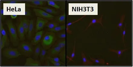
ICC/IF analysis of HeLa cells and negative control NIH3T3 cells using GTX22790 HSP27 antibody [G3.1]. Green : Primary antibody Blue : Nuclei Red : Actin Fixation : Formalin Permeabilization : 0.1% Triton X-100 in TBS for 10 minutes Dilution : 1:50 for at least 1 hour at room temperature
![ICC/IF analysis of U251 cells using GTX22790 HSP27 antibody [G3.1]. Cells were probed without (right) or with(left) an antibody. Green : Primary antibody Blue : Nuclei Red : Actin Fixation : formaldehyde Dilution : 1:200 overnight at 4oC](https://www.genetex.com/upload/website/prouct_img/normal/GTX22790/GTX22790_450_ICC-IF_w_23060620_628.webp "ICC/IF analysis of U251 cells using GTX22790 HSP27 antibody [G3.1]. Cells were probed without (right) or with(left) an antibody. Green : Primary antibody Blue : Nuclei Red : Actin Fixation : formaldehyde Dilution : 1:200 overnight at 4oC")
![IHC-P analysis of human tonsil tissue using GTX22790 HSP27 antibody [G3.1]. Left : Primary antibody Right : Negative control without primary antibody Antigen retrieval : heat induced antigen retrieval was performed using 10mM sodium citrate (pH6.0) buffer, microwaved for 8-15 minutes Dilution : 1:500](https://www.genetex.com/upload/website/prouct_img/normal/GTX22790/GTX22790_1115_IHC-P_w_23060620_862.webp "IHC-P analysis of human tonsil tissue using GTX22790 HSP27 antibody [G3.1]. Left : Primary antibody Right : Negative control without primary antibody Antigen retrieval : heat induced antigen retrieval was performed using 10mM sodium citrate (pH6.0) buffer, microwaved for 8-15 minutes Dilution : 1:500")
![ICC/IF analysis of HeLa cells using GTX22790 HSP27 antibody [G3.1]. Cells were probed without (right) or with(left) an antibody. Green : Primary antibody Blue : Nuclei Red : Actin Fixation : formaldehyde Dilution : 1:200 overnight at 4oC](https://www.genetex.com/upload/website/prouct_img/normal/GTX22790/GTX22790_449_ICC-IF_w_23060620_998.webp "ICC/IF analysis of HeLa cells using GTX22790 HSP27 antibody [G3.1]. Cells were probed without (right) or with(left) an antibody. Green : Primary antibody Blue : Nuclei Red : Actin Fixation : formaldehyde Dilution : 1:200 overnight at 4oC")
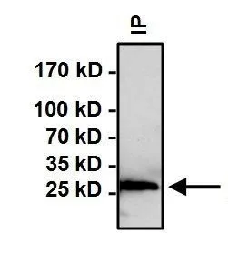
![IP analysis of HeLa cell lysates using GTX22790 HSP27 antibody [G3.1]. GTX22790 was used as IP and IB antibody IP reaction : 2μg antibody / 500μg lysate WB antibody dilution : 1:1000](https://www.genetex.com/upload/website/prouct_img/normal/GTX22790/GTX22790_1433_IP_w_23060620_847.webp "IP analysis of HeLa cell lysates using GTX22790 HSP27 antibody [G3.1]. GTX22790 was used as IP and IB antibody IP reaction : 2μg antibody / 500μg lysate WB antibody dilution : 1:1000")
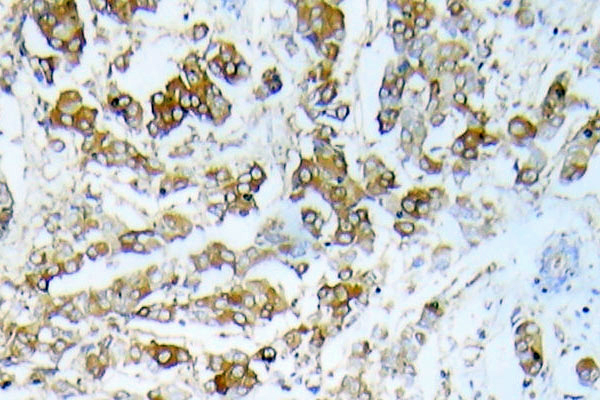
![IHC-P analysis of human breast carcinoma tissue using GTX22790 HSP27 antibody [G3.1]. Left : Primary antibody Right : Negative control without primary antibody Antigen retrieval : heat induced antigen retrieval was performed using 10mM sodium citrate (pH6.0) buffer, microwaved for 8-15 minutes Dilution : 1:500](https://www.genetex.com/upload/website/prouct_img/normal/GTX22790/GTX22790_1113_IHC-P_w_23060620_380.webp "IHC-P analysis of human breast carcinoma tissue using GTX22790 HSP27 antibody [G3.1]. Left : Primary antibody Right : Negative control without primary antibody Antigen retrieval : heat induced antigen retrieval was performed using 10mM sodium citrate (pH6.0) buffer, microwaved for 8-15 minutes Dilution : 1:500")
![ICC/IF analysis of C6 cells using GTX22790 HSP27 antibody [G3.1]. Cells were probed without (right) or with(left) an antibody. Green : Primary antibody Blue : Nuclei Red : Actin Fixation : formaldehyde Dilution : 1:20 overnight at 4oC](https://www.genetex.com/upload/website/prouct_img/normal/GTX22790/GTX22790_448_ICC-IF_w_23060620_469.webp "ICC/IF analysis of C6 cells using GTX22790 HSP27 antibody [G3.1]. Cells were probed without (right) or with(left) an antibody. Green : Primary antibody Blue : Nuclei Red : Actin Fixation : formaldehyde Dilution : 1:20 overnight at 4oC")
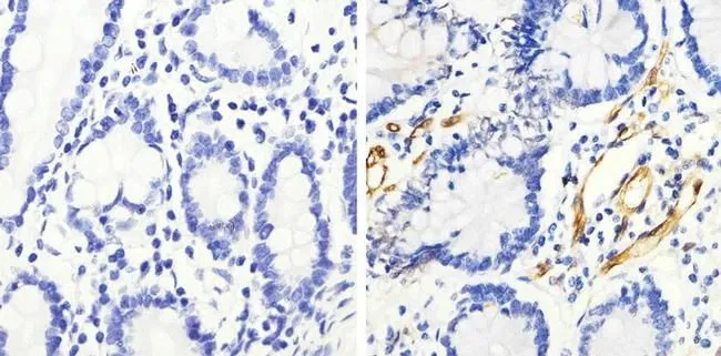
![IHC-P analysis of human colon carcinoma tissue using GTX22790 HSP27 antibody [G3.1]. Left : Primary antibody Right : Negative control without primary antibody Antigen retrieval : heat induced antigen retrieval was performed using 10mM sodium citrate (pH6.0) buffer, microwaved for 8-15 minutes Dilution : 1:200](https://www.genetex.com/upload/website/prouct_img/normal/GTX22790/GTX22790_1114_IHC-P_w_23060620_186.webp "IHC-P analysis of human colon carcinoma tissue using GTX22790 HSP27 antibody [G3.1]. Left : Primary antibody Right : Negative control without primary antibody Antigen retrieval : heat induced antigen retrieval was performed using 10mM sodium citrate (pH6.0) buffer, microwaved for 8-15 minutes Dilution : 1:200")
![WB analysis of 50μg of the indicated whole cell lysates using GTX22790 HSP27 antibody [G3.1]. Dilution : 1:1000](https://www.genetex.com/upload/website/prouct_img/normal/GTX22790/GTX22790_1594_WB_w_23060620_922.webp "WB analysis of 50μg of the indicated whole cell lysates using GTX22790 HSP27 antibody [G3.1]. Dilution : 1:1000")
![ICC/IF analysis of HeLa cells using GTX22790 HSP27 antibody [G3.1]. Cells were probed without (right) or with(left) an antibody. Green : Primary antibody Blue : Nuclei Red : Actin Fixation : Formalin Permeabilization : 0.1% Triton X-100 in TBS for 10 minutes Dilution : 1:100 for at least 1 hour at room temperature](https://www.genetex.com/upload/website/prouct_img/normal/GTX22790/GTX22790_447_ICC-IF_w_23060620_477.webp "ICC/IF analysis of HeLa cells using GTX22790 HSP27 antibody [G3.1]. Cells were probed without (right) or with(left) an antibody. Green : Primary antibody Blue : Nuclei Red : Actin Fixation : Formalin Permeabilization : 0.1% Triton X-100 in TBS for 10 minutes Dilution : 1:100 for at least 1 hour at room temperature")
ICC/IF analysis of HeLa cells and negative control NIH3T3 cells using GTX22790 HSP27 antibody [G3.1]. Green : Primary antibody Blue : Nuclei Red : Actin Fixation : Formalin Permeabilization : 0.1% Triton X-100 in TBS for 10 minutes Dilution : 1:50 for at least 1 hour at room temperature
HSP27 antibody [G3.1]
GTX22790
ApplicationsImmunoFluorescence, ImmunoPrecipitation, Western Blot, ELISA, ImmunoCytoChemistry, ImmunoHistoChemistry, ImmunoHistoChemistry Paraffin, Other Application
Product group Antibodies
ReactivityCanine, Human, Mouse, Primate, Rat
TargetHSPB1
Overview
- SupplierGeneTex
- Product NameHSP27 antibody [G3.1]
- Delivery Days Customer9
- Application Supplier NoteWB: 1:500 - 1:1000. ICC/IF: 1:50 - 1:500. IHC-P: 1:500. IP: 2 microl. *Optimal dilutions/concentrations should be determined by the researcher.Not tested in other applications.
- ApplicationsImmunoFluorescence, ImmunoPrecipitation, Western Blot, ELISA, ImmunoCytoChemistry, ImmunoHistoChemistry, ImmunoHistoChemistry Paraffin, Other Application
- CertificationResearch Use Only
- ClonalityMonoclonal
- Clone IDG3.1
- ConjugateUnconjugated
- Gene ID3315
- Target nameHSPB1
- Target descriptionheat shock protein family B (small) member 1
- Target synonymsCMT2F, HEL-S-102, HMN2B, HMND3, HS.76067, HSP27, HSP28, Hsp25, SRP27, heat shock protein beta-1, 28 kDa heat shock protein, epididymis secretory protein Li 102, estrogen-regulated 24 kDa protein, heat shock 27 kDa protein, heat shock 27kD protein 1, heat shock 27kDa protein 1, heat shock protein family B member 1, stress-responsive protein 27
- HostMouse
- IsotypeIgG1
- Protein IDP04792
- Protein NameHeat shock protein beta-1
- Scientific DescriptionThis gene encodes a member of the small heat shock protein (HSP20) family of proteins. In response to environmental stress, the encoded protein translocates from the cytoplasm to the nucleus and functions as a molecular chaperone that promotes the correct folding of other proteins. This protein plays an important role in the differentiation of a wide variety of cell types. Expression of this gene is correlated with poor clinical outcome in multiple human cancers, and the encoded protein may promote cancer cell proliferation and metastasis, while protecting cancer cells from apoptosis. Mutations in this gene have been identified in human patients with Charcot-Marie-Tooth disease and distal hereditary motor neuropathy. [provided by RefSeq, Aug 2017]
- ReactivityCanine, Human, Mouse, Primate, Rat
- Storage Instruction-20°C or -80°C,2°C to 8°C
- UNSPSC12352203
Datasheet
Related products
Product group Antibodies

Anti-HSPB1 [SAIC-23B-22]AB00313-1.1-BT
ApplicationsMass Spectrometry
ReactivityHuman
TargetHSPB1
- SizePrice
Product group Antibodies
Anti-HSP27 Antibody130-10055
ApplicationsELISA
ReactivityHuman
TargetHSPB1
- SizePrice
Product group Antibodies
HSPB1 Polyclonal AntibodyCAC15125
ApplicationsWestern Blot, ELISA, ImmunoHistoChemistry
TargetHSPB1
- SizePrice
Product group Antibodies
References
HSP27 Polyclonal AntibodyBS-0730R
ApplicationsFlow Cytometry, ImmunoFluorescence, Western Blot, ELISA, ImmunoCytoChemistry, ImmunoHistoChemistry, ImmunoHistoChemistry Frozen, ImmunoHistoChemistry Paraffin
ReactivityBovine, Canine, Human, Mouse, Porcine, Rat
TargetHSPB1
- SizePrice
Product group Antibodies
HSPB1 Monoclonal AntibodyCSB-MA080263
ApplicationsWestern Blot, ELISA, ImmunoHistoChemistry
ReactivityHuman
TargetHSPB1
- SizePrice
Product group Antibodies
Anti-HSP27 AntibodyA100672
ApplicationsELISA, ImmunoHistoChemistry
ReactivityHuman
- SizePrice
Product group Antibodies
HSP27 (phospho Ser82) antibodyGTX17677
ApplicationsImmunoFluorescence, Western Blot, ELISA, ImmunoCytoChemistry, ImmunoHistoChemistry, ImmunoHistoChemistry Paraffin
ReactivityHuman, Mouse, Rat
TargetHSPB1
- SizePrice
Product group Antibodies
HSP27 (phospho Ser82) antibodyGTX17937
ApplicationsFlow Cytometry, Western Blot, ImmunoHistoChemistry, ImmunoHistoChemistry Paraffin
ReactivityHuman
TargetHSPB1
- SizePrice
Product group Antibodies
HSP27 (phospho Ser15) antibodyGTX25581
ApplicationsFlow Cytometry, ImmunoFluorescence, ImmunoPrecipitation, Western Blot, ImmunoCytoChemistry, ImmunoHistoChemistry
ReactivityHuman, Rabbit, Rat
TargetHSPB1
- SizePrice