

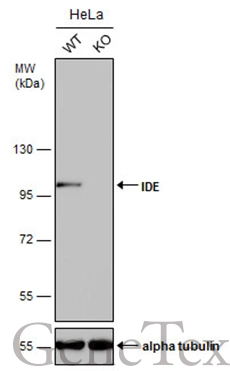
Wild-type (WT) and IDE knockout (KO) HeLa cell extracts (30 μg) were separated by 7.5% SDS-PAGE, and the membrane was blotted with IDE antibody [GT143] (GTX633694) diluted at 1:1000. The HRP-conjugated anti-mouse IgG antibody (GTX213111-01) was used to detect the primary antibody.
![IDE antibody [GT143] detects IDE protein at cytoplasm in mouse pancreas by immunohistochemical analysis. Sample: Paraffin-embedded mouse pancreas. IDE antibody [GT143] (GTX633694) diluted at 1:200.
Antigen Retrieval: Citrate buffer, pH 6.0, 15 min](https://www.genetex.com/upload/website/prouct_img/normal/GTX633694/GTX633694_42618_20161219_IHC-P_M_w_23061202_126.webp "IDE antibody [GT143] detects IDE protein at cytoplasm in mouse pancreas by immunohistochemical analysis. Sample: Paraffin-embedded mouse pancreas. IDE antibody [GT143] (GTX633694) diluted at 1:200.
Antigen Retrieval: Citrate buffer, pH 6.0, 15 min")
![IDE antibody [GT143] detects IDE protein at cytoplasm in rat colon by immunohistochemical analysis. Sample: Paraffin-embedded rat colon. IDE antibody [GT143] (GTX633694) diluted at 1:200.
Antigen Retrieval: Citrate buffer, pH 6.0, 15 min](https://www.genetex.com/upload/website/prouct_img/normal/GTX633694/GTX633694_42618_20161219_IHC-P_R_w_23061202_344.webp "IDE antibody [GT143] detects IDE protein at cytoplasm in rat colon by immunohistochemical analysis. Sample: Paraffin-embedded rat colon. IDE antibody [GT143] (GTX633694) diluted at 1:200.
Antigen Retrieval: Citrate buffer, pH 6.0, 15 min")
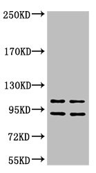
![Various tissue extracts (50 μg) were separated by 7.5% SDS-PAGE, and the membrane was blotted with IDE antibody [GT143] (GTX633694) diluted at 1:1000. The HRP-conjugated anti-mouse IgG antibody (GTX213111-01) was used to detect the primary antibody.](https://www.genetex.com/upload/website/prouct_img/normal/GTX633694/GTX633694_42618_20170615_WB_M_R_w_23061202_730.webp "Various tissue extracts (50 μg) were separated by 7.5% SDS-PAGE, and the membrane was blotted with IDE antibody [GT143] (GTX633694) diluted at 1:1000. The HRP-conjugated anti-mouse IgG antibody (GTX213111-01) was used to detect the primary antibody.")
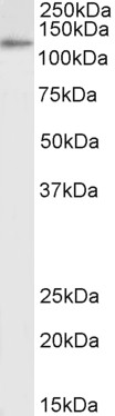
![Whole cell extract (30 μg) was separated by 7.5% SDS-PAGE, and the membrane was blotted with IDE antibody [GT143] (GTX633694) diluted at 1:1000.](https://www.genetex.com/upload/website/prouct_img/normal/GTX633694/GTX633694_42618_20160929_WB_w_23061202_487.webp "Whole cell extract (30 μg) was separated by 7.5% SDS-PAGE, and the membrane was blotted with IDE antibody [GT143] (GTX633694) diluted at 1:1000.")
Wild-type (WT) and IDE knockout (KO) HeLa cell extracts (30 μg) were separated by 7.5% SDS-PAGE, and the membrane was blotted with IDE antibody [GT143] (GTX633694) diluted at 1:1000. The HRP-conjugated anti-mouse IgG antibody (GTX213111-01) was used to detect the primary antibody.
IDE antibody [GT143]
GTX633694
ApplicationsWestern Blot, ImmunoHistoChemistry, ImmunoHistoChemistry Paraffin
Product group Antibodies
ReactivityHuman, Mouse, Rat
TargetIDE
Overview
- SupplierGeneTex
- Product NameIDE antibody [GT143]
- Delivery Days Customer9
- Application Supplier NoteWB: 1:500-1:3000. IHC-P: 1:100-1:1000. *Optimal dilutions/concentrations should be determined by the researcher.Not tested in other applications.
- ApplicationsWestern Blot, ImmunoHistoChemistry, ImmunoHistoChemistry Paraffin
- CertificationResearch Use Only
- ClonalityMonoclonal
- Clone IDGT143
- Concentration1 mg/ml
- ConjugateUnconjugated
- Gene ID3416
- Target nameIDE
- Target descriptioninsulin degrading enzyme
- Target synonymsINSULYSIN, insulin-degrading enzyme, Abeta-degrading protease, insulin protease, insulinase
- HostMouse
- IsotypeIgG2b
- Protein IDP14735
- Protein NameInsulin-degrading enzyme
- Scientific DescriptionThis gene may belong to a protease family responsible for intercellular peptide signalling. Though its role in the cellular processing of insulin has not yet been defined, insulin-degrading enzyme is thought to be involved in the termination of the insulin response. [provided by RefSeq]
- ReactivityHuman, Mouse, Rat
- Storage Instruction-20°C or -80°C,2°C to 8°C
- UNSPSC41116161
Datasheet
Related products
Product group Antibodies
Anti-IDE Antibody144-01630
ApplicationsImmunoFluorescence, Western Blot, ImmunoHistoChemistry
ReactivityHuman, Mouse
TargetIDE
- SizePrice
Product group Antibodies
ApplicationsWestern Blot, ELISA
ReactivityHuman
- SizePrice
Product group Antibodies

Insulysin AntibodyABX430884
ApplicationsWestern Blot, ELISA, ImmunoHistoChemistry
- SizePrice
Product group Antibodies

Anti-Insulin-degrading Enzyme [Fab-IDE]AB00406-1.1-BT
ApplicationsELISA
ReactivityHuman
TargetIDE
- SizePrice

Product group Antibodies
ApplicationsWestern Blot, ImmunoHistoChemistry
ReactivityHuman, Mouse, Rat
TargetIDE
- SizePrice
Product group Antibodies

IDE Antibody (clone 1H4)LS-C767393
ApplicationsImmunoFluorescence, Western Blot, ImmunoHistoChemistry, ImmunoHistoChemistry Paraffin
ReactivityHuman
TargetIDE
- SizePrice
Product group Antibodies
TargetIDE
- SizePrice
Product group Antibodies

ApplicationsWestern Blot, ELISA, ImmunoHistoChemistry
ReactivityCanine, Human
TargetIDE
- SizePrice

Product group Antibodies
Anti-IDE AntibodyHPA063478
ApplicationsWestern Blot, ImmunoHistoChemistry
ReactivityHuman
TargetIDE
- SizePrice
Product group Antibodies
IDE Monoclonal AntibodyCSB-MA000241
ApplicationsWestern Blot, ELISA
ReactivityHuman
TargetIDE
- SizePrice