

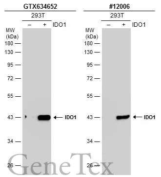
Non-transfected (–) and transfected (+) 293T whole cell extracts (30 μg) were separated by 10% SDS-PAGE, and the membranes were blotted with IDO1 antibody [GT273] (GTX634652) diluted at 1:5000 and competitor's antibody (CST#12006) diluted at 1:1000. The HRP-conjugated anti-mouse IgG antibody (GTX213111-01) was used to detect the primary antibody. *The competitor is not affiliated with GeneTex and does not endorse this product.
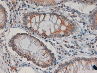
![IDO1 antibody [GT273] detects IDO1 protein at cell membrane and cytoplasm by immunohistochemical analysis. Sample: Paraffin-embedded human tonsil. IDO1 stained by IDO1 antibody [GT273] (GTX634652) diluted at 1:200. Antigen Retrieval: Citrate buffer, pH 6.0, 15 min](https://www.genetex.com/upload/website/prouct_img/normal/GTX634652/GTX634652_44524_20220121_IHC-P_w_23061202_761.webp "IDO1 antibody [GT273] detects IDO1 protein at cell membrane and cytoplasm by immunohistochemical analysis. Sample: Paraffin-embedded human tonsil. IDO1 stained by IDO1 antibody [GT273] (GTX634652) diluted at 1:200. Antigen Retrieval: Citrate buffer, pH 6.0, 15 min")
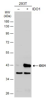
![Untreated (–) and treated (+) A549 whole cell extracts (30 μg) were separated by 10% SDS-PAGE, and the membrane was blotted with IDO1 antibody [GT273] (GTX634652) diluted at 1:5000. The HRP-conjugated anti-mouse IgG antibody (GTX213111-01) was used to detect the primary antibody.](https://www.genetex.com/upload/website/prouct_img/normal/GTX634652/GTX634652_44524_20211210_WB_treatment_IFN-gamma_w_23061202_391.webp "Untreated (–) and treated (+) A549 whole cell extracts (30 μg) were separated by 10% SDS-PAGE, and the membrane was blotted with IDO1 antibody [GT273] (GTX634652) diluted at 1:5000. The HRP-conjugated anti-mouse IgG antibody (GTX213111-01) was used to detect the primary antibody.")
Non-transfected (–) and transfected (+) 293T whole cell extracts (30 μg) were separated by 10% SDS-PAGE, and the membranes were blotted with IDO1 antibody [GT273] (GTX634652) diluted at 1:5000 and competitor's antibody (CST#12006) diluted at 1:1000. The HRP-conjugated anti-mouse IgG antibody (GTX213111-01) was used to detect the primary antibody. *The competitor is not affiliated with GeneTex and does not endorse this product.
IDO1 antibody [GT273]
GTX634652
ApplicationsWestern Blot, ImmunoHistoChemistry, ImmunoHistoChemistry Paraffin
Product group Antibodies
ReactivityHuman, Mouse
TargetIDO1
Overview
- SupplierGeneTex
- Product NameIDO1 antibody [GT273]
- Delivery Days Customer9
- Application Supplier NoteWB: 1:1000-1:10000. IHC-P: 1:100-1:1000. *Optimal dilutions/concentrations should be determined by the researcher.Not tested in other applications.
- ApplicationsWestern Blot, ImmunoHistoChemistry, ImmunoHistoChemistry Paraffin
- CertificationResearch Use Only
- ClonalityMonoclonal
- Clone IDGT273
- Concentration1 mg/ml
- ConjugateUnconjugated
- Gene ID3620
- Target nameIDO1
- Target descriptionindoleamine 2,3-dioxygenase 1
- Target synonymsIDO, IDO-1, INDO, indoleamine 2,3-dioxygenase 1, indolamine 2,3 dioxygenase, indole 2,3-dioxygenase, indoleamine-pyrrole 2,3-dioxygenase
- HostMouse
- IsotypeIgG2a
- Protein IDP14902
- Protein NameIndoleamine 2,3-dioxygenase 1
- Scientific DescriptionThis gene encodes indoleamine 2,3-dioxygenase (IDO) - a heme enzyme that catalyzes the first and rate-limiting step in tryptophan catabolism to N-formyl-kynurenine. This enzyme acts on multiple tryptophan substrates including D-tryptophan, L-tryptophan, 5-hydroxy-tryptophan, tryptamine, and serotonin. This enzyme is thought to play a role in a variety of pathophysiological processes such as antimicrobial and antitumor defense, neuropathology, immunoregulation, and antioxidant activity. Through its expression in dendritic cells, monocytes, and macrophages this enzyme modulates T-cell behavior by its peri-cellular catabolization of the essential amino acid tryptophan.[provided by RefSeq, Feb 2011]
- ReactivityHuman, Mouse
- Storage Instruction-20°C or -80°C,2°C to 8°C
- UNSPSC12352203
References
- Xu C, Feng C, Huang P, et al. TNFα and IFNγ rapidly activate PI3K-AKT signaling to drive glycolysis that confers mesenchymal stem cells enhanced anti-inflammatory property. Stem Cell Res Ther. 2022,13(1):491. doi: 10.1186/s13287-022-03178-3Read this paper
- Sorrentino C, D'Antonio L, Fieni C, et al. Colorectal Cancer-Associated Immune Exhaustion Involves T and B Lymphocytes and Conventional NK Cells and Correlates With a Shorter Overall Survival. Front Immunol. 2021,12:778329. doi: 10.3389/fimmu.2021.778329Read this paper
Datasheet
Related products
Product group Antibodies

Anti-IDO1 [19C5]Ab03196-10.0
ApplicationsELISA, ImmunoHistoChemistry
ReactivityHuman
TargetIDO1
- SizePrice
Product group Antibodies
Anti-IDO1 Antibody144-64139
ApplicationsWestern Blot
ReactivityHuman, Mouse
TargetIDO1
- SizePrice
Product group Antibodies
References
IDO1 antibodyGTX113753
ApplicationsWestern Blot, ImmunoHistoChemistry, ImmunoHistoChemistry Frozen
ReactivityHuman, Rat
TargetIDO1
- SizePrice
Product group Antibodies
Ido1 Polyclonal AntibodyCAC08537
ApplicationsImmunoFluorescence, Western Blot, ELISA, ImmunoHistoChemistry
TargetIDO1
- SizePrice
Product group Antibodies
References
IDO1 Polyclonal AntibodyBS-15493R
ApplicationsFlow Cytometry, ImmunoFluorescence, Western Blot, ELISA, ImmunoCytoChemistry, ImmunoHistoChemistry, ImmunoHistoChemistry Frozen, ImmunoHistoChemistry Paraffin
ReactivityHuman, Mouse, Rat
TargetIDO1
- SizePrice
Product group Antibodies
IDO1 antibody, InternalGTX89419
ApplicationsWestern Blot, ImmunoHistoChemistry, ImmunoHistoChemistry Paraffin
ReactivityHuman
TargetIDO1
- SizePrice
Product group Antibodies
anti-IDO1 (human), pAbAG-25A-0029
ApplicationsFlow Cytometry, Western Blot, ELISA, ImmunoCytoChemistry
ReactivityHuman
TargetIDO1
- SizePrice
Product group Antibodies
References
IDO1 antibody [10.1]GTX54075
ApplicationsWestern Blot, ImmunoHistoChemistry, ImmunoHistoChemistry Frozen
ReactivityHuman, Mouse
TargetIDO1
- SizePrice