

IGF2R (Phospho-Ser2409) Antibody
ABX012519
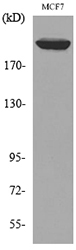
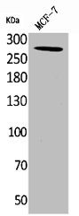
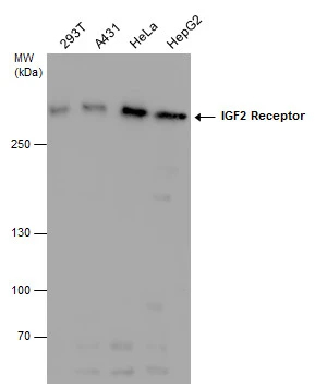
ApplicationsWestern Blot, ELISA, ImmunoHistoChemistry
Product group Antibodies
Overview
- SupplierAbbexa
- Product NameIGF2R (Phospho-Ser2409) Antibody
- Delivery Days Customer12
- ApplicationsWestern Blot, ELISA, ImmunoHistoChemistry
- CertificationResearch Use Only
- ClonalityPolyclonal
- ConjugateUnconjugated
- HostRabbit
- UNSPSC12352203
Related products
Product group Antibodies
Anti-IGF2R AntibodyA101029
ApplicationsWestern Blot, ELISA
ReactivityHuman
- SizePrice

Product group Antibodies
Anti-Mannose 6 Phosphate Receptor (Cation independent)/IGF2R Antibody Picoband(r)A00951-CARRIER-FREE
ApplicationsFlow Cytometry, ImmunoFluorescence, Western Blot, ELISA, ImmunoCytoChemistry, ImmunoHistoChemistry
ReactivityHuman, Mouse, Rat
TargetIGF2R
- SizePrice
Product group Antibodies
Anti-IGF2R Antibody144-60151
ApplicationsImmunoFluorescence, Western Blot, ImmunoHistoChemistry
ReactivityHuman, Mouse, Rat
TargetIGF2R
- SizePrice
Product group Antibodies
IGF2R/M6PR Polyclonal AntibodyBS-6670R
ApplicationsFlow Cytometry, ImmunoFluorescence, ELISA, ImmunoCytoChemistry, ImmunoHistoChemistry, ImmunoHistoChemistry Frozen, ImmunoHistoChemistry Paraffin
ReactivityBovine, Canine, Chicken, Equine, Human, Mouse, Porcine, Rabbit, Rat
TargetIGF2R
- SizePrice
Product group Antibodies
ApplicationsImmunoPrecipitation, Western Blot, ImmunoCytoChemistry, ImmunoHistoChemistry
TargetIGF2R
- SizePrice
Product group Antibodies
IGF2R AntibodyCSB-PA005283
ApplicationsWestern Blot, ELISA
ReactivityHuman
TargetIGF2R
- SizePrice
Product group Antibodies

IGF2R / CD222 AntibodyLS-C406473
ApplicationsELISA, ImmunoHistoChemistry
ReactivityHuman, Mouse
TargetIGF2R
- SizePrice

Product group Antibodies
Anti-IGF2R AntibodyHPA011332
ApplicationsImmunoCytoChemistry, ImmunoHistoChemistry
ReactivityHuman
TargetIGF2R
- SizePrice
Product group Antibodies
IGF2R antibodyGTX130109
ApplicationsWestern Blot
ReactivityHuman
TargetIGF2R
- SizePrice