

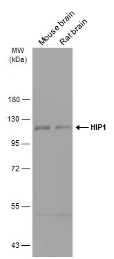
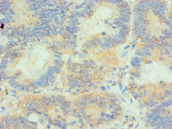
IHC-plus(tm) HIP1 Antibody
LS-B14586
ApplicationsImmunoFluorescence, Western Blot, ImmunoCytoChemistry, ImmunoHistoChemistry, ImmunoHistoChemistry Paraffin
Product group Antibodies
ReactivityHuman, Mouse
TargetHIP1
Overview
- SupplierLifeSpan BioSciences
- Product NameIHC-plus(tm) HIP1 Antibody
- Delivery Days Customer23
- ApplicationsImmunoFluorescence, Western Blot, ImmunoCytoChemistry, ImmunoHistoChemistry, ImmunoHistoChemistry Paraffin
- Applications SupplierICC (1:50 - 1:100), IF (1:50 - 1:100), IHC, IHC-P (1:50 - 1:200), WB (1:500 - 1:2000)
- CertificationResearch Use Only
- ClonalityPolyclonal
- Concentration1 mg/ml
- ConjugateUnconjugated
- Estimated Purity...
- Gene ID3092
- Target nameHIP1
- Target descriptionhuntingtin interacting protein 1
- Target synonymsHIP-I, ILWEQ, SHON, SHONbeta, SHONgamma, huntingtin-interacting protein 1, huntingtin-interacting protein I
- HostRabbit
- ReactivityHuman, Mouse
- Storage Instruction-20°C
- UNSPSC12352203
Related products
Product group Antibodies
Anti-HIP1 Antibody144-06921
ApplicationsImmunoFluorescence, Western Blot, ImmunoHistoChemistry
ReactivityHuman, Mouse
TargetHIP1
- SizePrice

Product group Antibodies
References
ApplicationsImmunoFluorescence, ImmunoPrecipitation, Western Blot, ImmunoCytoChemistry
ReactivityHuman, Mouse, Rat
TargetHIP1
- SizePrice
Product group Antibodies
HIP1 antibodyGTX131449
ApplicationsWestern Blot
ReactivityHuman, Mouse, Rat
TargetHIP1
- SizePrice
Product group Antibodies
HIP1 Recombinant Antibody, AbBy Fluor-594 ConjugatedBSM-61799R-BF594
ApplicationsImmunoFluorescence, Western Blot, ImmunoCytoChemistry
ReactivityHuman, Mouse, Rat
TargetHIP1
- SizePrice
Product group Antibodies
HIP1 AntibodyCSB-PA010365ESR1HU
ApplicationsELISA, ImmunoHistoChemistry
ReactivityHuman
TargetHIP1
- SizePrice
Product group Antibodies
Anti-HIP1 AntibodyA31749
ApplicationsImmunoFluorescence, Western Blot, ImmunoHistoChemistry
ReactivityHuman, Mouse
- SizePrice

Product group Antibodies
Anti-HIP1 AntibodyHPA013606
ApplicationsWestern Blot, ImmunoCytoChemistry, ImmunoHistoChemistry
ReactivityHuman, Mouse, Rat
TargetHIP1
- SizePrice