

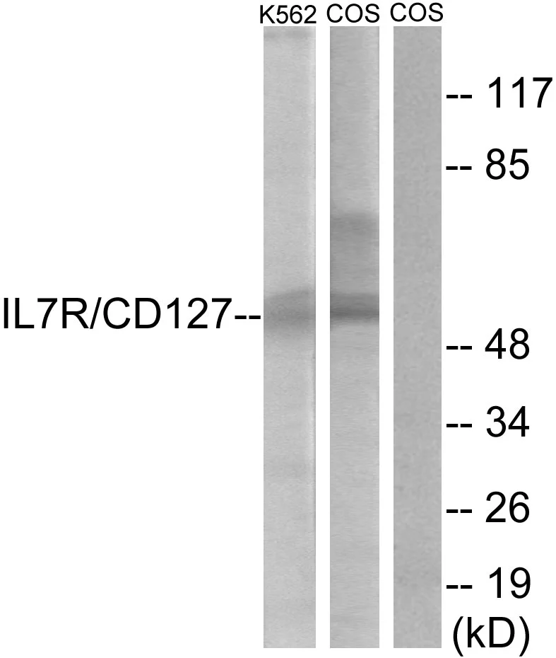
WB analysis of K562 and COS cells treated with insulin 0.01U/ml (15mins) lysate using GTX87162 IL7 Receptor alpha antibody. The lane on the right is blocked with the synthesized peptide.
WB analysis of K562 and COS cells treated with insulin 0.01U/ml (15mins) lysate using GTX87162 IL7 Receptor alpha antibody. The lane on the right is blocked with the synthesized peptide.
IL7 Receptor alpha antibody
GTX87162
ApplicationsWestern Blot
Product group Antibodies
TargetIL7R
Overview
- SupplierGeneTex
- Product NameIL7 Receptor alpha antibody
- Delivery Days Customer9
- Application Supplier NoteWB: 1:500~1:1000. *Optimal dilutions/concentrations should be determined by the researcher.Not tested in other applications.
- ApplicationsWestern Blot
- CertificationResearch Use Only
- ClonalityPolyclonal
- ConjugateUnconjugated
- Gene ID3575
- Target nameIL7R
- Target descriptioninterleukin 7 receptor
- Target synonymsCD127, CDW127, IL-7R-alpha, IL-7Ralpha, IL7RA, IL7Ralpha, ILRA, IMD104, lnc-IL7R, sIL-7R, interleukin-7 receptor subunit alpha, CD127 antigen, IL-7 receptor subunit alpha, IL-7R subunit alpha, interleukin 7 receptor alpha chain, soluble interleukin-7 receptor
- HostRabbit
- IsotypeIgG
- Protein IDP16871
- Protein NameInterleukin-7 receptor subunit alpha
- Scientific DescriptionThe protein encoded by this gene is a receptor for interleukine 7 (IL7). The function of this receptor requires the interleukin 2 receptor, gamma chain (IL2RG), which is a common gamma chain shared by the receptors of various cytokines, including interleukine 2, 4, 7, 9, and 15. This protein has been shown to play a critical role in the V(D)J recombination during lymphocyte development. This protein is also found to control the accessibility of the TCR gamma locus by STAT5 and histone acetylation. Knockout studies in mice suggested that blocking apoptosis is an essential function of this protein during differentiation and activation of T lymphocytes. The functional defects in this protein may be associated with the pathogenesis of the severe combined immunodeficiency (SCID). Alternatively spliced transcript variants have been found for this gene. [provided by RefSeq, May 2014]
- Storage Instruction-20°C or -80°C,2°C to 8°C
- UNSPSC12352203
Datasheet
Related products
Product group Antibodies
IL7R AntibodyCSB-PA003034
ApplicationsWestern Blot, ELISA
ReactivityHuman, Monkey, Mouse
TargetIL7R
- SizePrice
Product group Antibodies
IL7 Receptor alpha antibodyGTX54311
ApplicationsImmunoFluorescence, Western Blot, ImmunoCytoChemistry
TargetIL7R
- SizePrice
Product group Antibodies
ApplicationsWestern Blot
TargetIL7R
- SizePrice
Product group Antibodies
ApplicationsImmunoPrecipitation, Western Blot, ImmunoHistoChemistry
TargetIL7R
- SizePrice
Product group Antibodies

Anti-CD127 [1A11]Ab02640-1.1
ApplicationsWestern Blot, ELISA, ImmunoHistoChemistry, Other Application
TargetIL7R
- SizePrice
Product group Antibodies
Anti-CD127 Antibody144-62283
ApplicationsImmunoFluorescence, Western Blot
TargetIL7R
- SizePrice

Product group Antibodies
Anti-CD127/IL7R Antibody Picoband(r)A02222-3-CARRIER-FREE
ApplicationsFlow Cytometry, Western Blot, ELISA, ImmunoHistoChemistry
TargetIL7R
- SizePrice
Product group Antibodies
IL7R Polyclonal AntibodyCAC14850
ApplicationsImmunoFluorescence, Western Blot, ELISA
TargetIL7R
- SizePrice