

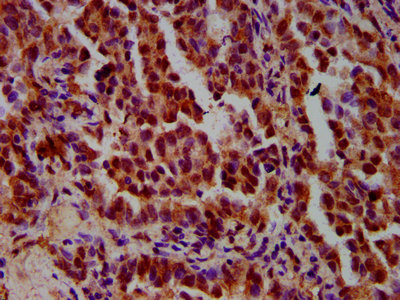
IHC image of CSB-PA22459A0Rb diluted at 1:600 and staining in paraffin-embedded human lung cancer performed on a Leica BondTM system. After dewaxing and hydration, antigen retrieval was mediated by high pressure in a citrate buffer (pH 6.0). Section was blocked with 10% normal goat serum 30min at RT. Then primary antibody (1% BSA) was incubated at 4°C overnight. The primary is detected by a biotinylated secondary antibody and visualized using an HRP conjugated SP system.
. Section was blocked with 10% normal goat serum 30min at RT. Then primary antibody (1% BSA) was incubated at 4°C overnight. The primary is detected by a biotinylated secondary antibody and visualized using an HRP conjugated SP system.")
.")
IHC image of CSB-PA22459A0Rb diluted at 1:600 and staining in paraffin-embedded human lung cancer performed on a Leica BondTM system. After dewaxing and hydration, antigen retrieval was mediated by high pressure in a citrate buffer (pH 6.0). Section was blocked with 10% normal goat serum 30min at RT. Then primary antibody (1% BSA) was incubated at 4°C overnight. The primary is detected by a biotinylated secondary antibody and visualized using an HRP conjugated SP system.
IMPDH1 Antibody
CSB-PA22459A0RB
ApplicationsImmunoFluorescence, ELISA, ImmunoHistoChemistry
Product group Antibodies
ReactivityHuman
TargetIMPDH1
Overview
- SupplierCusabio
- Product NameIMPDH1 Antibody
- Delivery Days Customer20
- ApplicationsImmunoFluorescence, ELISA, ImmunoHistoChemistry
- CertificationResearch Use Only
- ClonalityPolyclonal
- ConjugateUnconjugated
- Gene ID3614
- Target nameIMPDH1
- Target descriptioninosine monophosphate dehydrogenase 1
- Target synonymsIMPD, IMPD1, IMPDH-I, LCA11, RP10, sWSS2608, inosine-5'-monophosphate dehydrogenase 1, IMP (inosine 5'-monophosphate) dehydrogenase 1, IMP (inosine monophosphate) dehydrogenase 1, IMPD 1, IMPDH 1
- HostRabbit
- IsotypeIgG
- Protein IDP20839
- Protein NameInosine-5'-monophosphate dehydrogenase 1
- Scientific DescriptionCatalyzes the conversion of inosine 5-phosphate (IMP) to xanthosine 5-phosphate (XMP), the first committed and rate-limiting step in the de novo synthesis of guanine nucleotides, and therefore plays an important role in the regulation of cell growth. Could also have a single-stranded nucleic acid-binding activity and could play a role in RNA and/or DNA metabolism. It may also have a role in the development of malignancy and the growth progression of some tumors.
- ReactivityHuman
- Storage Instruction-20°C or -80°C
- UNSPSC41116161
Related products
Product group Antibodies
Anti-IMPDH1 AntibodyA16321
ApplicationsImmunoFluorescence, Western Blot, ImmunoCytoChemistry
ReactivityHuman, Mouse, Rat
- SizePrice

Product group Antibodies
Anti-IMPDH1 Antibody Picoband(r)A03791-1-CARRIER-FREE
ApplicationsFlow Cytometry, ImmunoFluorescence, Western Blot, ELISA, ImmunoCytoChemistry, ImmunoHistoChemistry
ReactivityHuman, Mouse
TargetIMPDH1
- SizePrice
Product group Antibodies

IMPDH1 AntibodyLS-C403248
ApplicationsWestern Blot, ELISA, ImmunoHistoChemistry
ReactivityHuman, Mouse, Rat
TargetIMPDH1
- SizePrice
Product group Antibodies
IMPDH1 Polyclonal AntibodyBS-6256R
ApplicationsImmunoFluorescence, Western Blot, ELISA, ImmunoCytoChemistry, ImmunoHistoChemistry, ImmunoHistoChemistry Frozen, ImmunoHistoChemistry Paraffin
ReactivityBovine, Canine, Guinea Pig, Human, Mouse, Porcine, Rabbit, Rat
TargetIMPDH1
- SizePrice
Product group Antibodies
IMPDH1 antibody [N1C1]GTX102814
ApplicationsWestern Blot
ReactivityHuman
TargetIMPDH1
- SizePrice
Product group Antibodies
Anti-IMPDH1 Antibody144-09497
ApplicationsWestern Blot
ReactivityHuman, Mouse, Rat
TargetIMPDH1
- SizePrice