

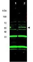
Western blot using GeneTex's purified anti-p47 ING3 antibody shows detection of a band at ~55 kDa corresponding to ING3 in RKO cells transfected with ING3 (lane 2). Control RKO cells do not show detection of this specific band (lane 1). The identity of the non-specific bands at 33 kDa and 20 kDa has not been determined. Each lane contains approximately 10 μg of RKO whole cell lysate separated on a 4-20% Tris-Glycine gel by SDS-PAGE and transferred to nitrocellulose. After blocking with 5% NF dry milk, the membrane was probed with the primary antibody diluted to 1:1,000. Incubation was at 4o C overnight followed by washes and reaction with a 1:20,000 dilution of IRDye?800 conjugated Rb-a-Goat IgG [H&L] MXHur for 45 min at room temperature. IRDye?800 fluorescence image was captured using the OdysseyR Infrared Imaging System developed by LI-COR. IRDye is a trademark of LI-COR, Inc. Other detection systems will yield similar results.

 and non-transfected (lane 1) RKO whole cell lysate using GTX48488 ING3 antibody. Dilution : 10 μg Dilution : 1:1000")
Western blot using GeneTex's purified anti-p47 ING3 antibody shows detection of a band at ~55 kDa corresponding to ING3 in RKO cells transfected with ING3 (lane 2). Control RKO cells do not show detection of this specific band (lane 1). The identity of the non-specific bands at 33 kDa and 20 kDa has not been determined. Each lane contains approximately 10 μg of RKO whole cell lysate separated on a 4-20% Tris-Glycine gel by SDS-PAGE and transferred to nitrocellulose. After blocking with 5% NF dry milk, the membrane was probed with the primary antibody diluted to 1:1,000. Incubation was at 4o C overnight followed by washes and reaction with a 1:20,000 dilution of IRDye?800 conjugated Rb-a-Goat IgG [H&L] MXHur for 45 min at room temperature. IRDye?800 fluorescence image was captured using the OdysseyR Infrared Imaging System developed by LI-COR. IRDye is a trademark of LI-COR, Inc. Other detection systems will yield similar results.
ING3 antibody
GTX48488
ApplicationsWestern Blot, ELISA
Product group Antibodies
ReactivityHuman
TargetING3
Overview
- SupplierGeneTex
- Product NameING3 antibody
- Delivery Days Customer9
- Application Supplier NoteWB: 1:200-1:2000. ELISA: 1:10000-1:40000. *Optimal dilutions/concentrations should be determined by the researcher.Not tested in other applications.
- ApplicationsWestern Blot, ELISA
- CertificationResearch Use Only
- ClonalityPolyclonal
- Concentration1.1 mg/ml
- ConjugateUnconjugated
- Gene ID54556
- Target nameING3
- Target descriptioninhibitor of growth family member 3
- Target synonymsEaf4, ING2, MEAF4, p47ING3, inhibitor of growth protein 3
- HostGoat
- IsotypeIgG
- Protein IDQ9NXR8
- Protein NameInhibitor of growth protein 3
- Scientific Descriptionp47 ING3 is a tumor suppressor protein similar to ING1 that can interact with TP53, inhibit cell growth, and induce apoptosis. This protein contains a PHD-finger, which is a common motif in proteins involved in chromatin remodeling. This gene can activate p53 trans-activated promoters, including promoters of p21/waf1 and bax. Over-expression of this gene has been shown to inhibit cell growth and induce apoptosis. Allelic loss and reduced expression of this gene were detected in head and neck cancers. Multiple alternatively spliced transcript variants have been observed.
- ReactivityHuman
- Storage Instruction-20°C or -80°C,2°C to 8°C
- UNSPSC41116161
Datasheet
Related products
Product group Antibodies
Anti-ING3 AntibodyA31079
ApplicationsImmunoFluorescence, ImmunoPrecipitation, Western Blot, ImmunoHistoChemistry
ReactivityHuman, Mouse, Rat
- SizePrice
Product group Antibodies
Anti-ING3 Antibody144-05832
ApplicationsImmunoFluorescence, Western Blot
ReactivityHuman, Mouse, Rat
TargetING3
- SizePrice
Product group Antibodies
ING3 AntibodyCSB-PA865171LA01HU
ApplicationsWestern Blot, ELISA, ImmunoHistoChemistry
ReactivityHuman, Mouse
TargetING3
- SizePrice
Product group Antibodies
ING3 Polyclonal AntibodyCAC14624
ApplicationsWestern Blot, ELISA, ImmunoHistoChemistry
ReactivityMouse
TargetING3
- SizePrice
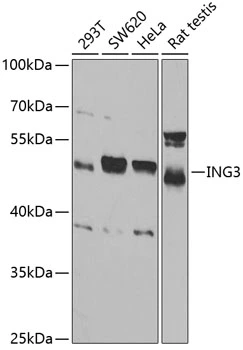
![Various whole cell extracts (30 μg) were separated by 10% SDS-PAGE, and the membrane was blotted with ING3 antibody [N2N3] (GTX102480) diluted at 1:1000. The HRP-conjugated anti-rabbit IgG antibody (GTX213110-01) was used to detect the primary antibody.](https://www.genetex.com/upload/website/prouct_img/normal/GTX102480/GTX102480_39617_20220908_WB_22091323_806.webp)
Product group Antibodies
ING3 antibody [N2N3]GTX102480
ApplicationsWestern Blot
ReactivityHuman
TargetING3
- SizePrice
Product group Antibodies

ING3 AntibodyLS-C334325
ApplicationsImmunoFluorescence, ImmunoPrecipitation, Western Blot, ImmunoHistoChemistry
ReactivityHuman, Mouse, Rat
TargetING3
- SizePrice

Product group Antibodies
Anti-ING3 AntibodyHPA067388
ApplicationsImmunoHistoChemistry
ReactivityHuman
TargetING3
- SizePrice
Product group Antibodies
ING3 antibodyGTX54356
ApplicationsImmunoFluorescence, Western Blot, ImmunoCytoChemistry
ReactivityHuman, Rat
TargetING3
- SizePrice
Product group Antibodies
Anti-ING3 AntibodyCAB5832
ApplicationsWestern Blot, ELISA
ReactivityHuman
TargetING3
- SizePrice