


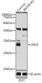
Western blot analysis is shown using GeneTex's Affinity Purified anti-p28 ING5 antibody to detect over expressed Human ING5 present in cell extracts. This western blot shows reactivity with purified recombinant human ING5 protein. Comparison to a molecular weight marker (not shown) indicates a single band of ~36 kDa corresponding to the expected molecular weight for the recombinant protein. Approximately 10 μg of lysate was separated on a 4-20% Tris-Glycine gel by SDS-PAGE and transferred onto nitrocellulose. After blocking the membrane was probed with the primary antibody diluted to 1:1,500.Incubation was overnight at 4o C followed by washes and reaction with a 1:20,000 dilution of IRDye?800 conjugated Rb-a-Goat IgG [H&L] MXHu for 45 min at room temperature. IRDye?800 fluorescence image was captured using the OdysseyR Infrared Imaging System developed by LI-COR. IRDye is a trademark of LI-COR, Inc. Other detection systems will yield similar results.
 and does not recognize TAP tagged ING4 on the same membrane (lane 2). A mock purification is shown in lane 1. Comparison to a molecular weight marker (not shown) indicates a single band of ~45.0 kDa corresponding to the expected molecular weight for the recombinant protein. The blot was incubated with a 1:500 dilution of the antibody at room temperature followed by detection using chemiluminescence reagent with a 5-min exposure time. Other detection systems will yield similar results.")


Western blot analysis is shown using GeneTex's Affinity Purified anti-p28 ING5 antibody to detect over expressed Human ING5 present in cell extracts. This western blot shows reactivity with purified recombinant human ING5 protein. Comparison to a molecular weight marker (not shown) indicates a single band of ~36 kDa corresponding to the expected molecular weight for the recombinant protein. Approximately 10 μg of lysate was separated on a 4-20% Tris-Glycine gel by SDS-PAGE and transferred onto nitrocellulose. After blocking the membrane was probed with the primary antibody diluted to 1:1,500.Incubation was overnight at 4o C followed by washes and reaction with a 1:20,000 dilution of IRDye?800 conjugated Rb-a-Goat IgG [H&L] MXHu for 45 min at room temperature. IRDye?800 fluorescence image was captured using the OdysseyR Infrared Imaging System developed by LI-COR. IRDye is a trademark of LI-COR, Inc. Other detection systems will yield similar results.
ING5 antibody
GTX48484
ApplicationsWestern Blot, ELISA
Product group Antibodies
ReactivityHuman
TargetING5
Overview
- SupplierGeneTex
- Product NameING5 antibody
- Delivery Days Customer9
- Application Supplier NoteWB: 1:500-1:1000. ELISA: 1:4000-1:16000. *Optimal dilutions/concentrations should be determined by the researcher.Not tested in other applications.
- ApplicationsWestern Blot, ELISA
- CertificationResearch Use Only
- ClonalityPolyclonal
- Concentration1.52 mg/ml
- ConjugateUnconjugated
- Gene ID84289
- Target nameING5
- Target descriptioninhibitor of growth family member 5
- Target synonymsp28ING5, inhibitor of growth protein 5
- HostGoat
- IsotypeIgG
- Protein IDQ8WYH8
- Protein NameInhibitor of growth protein 5
- Scientific Descriptionp28 ING5 is a tumor suppressor protein similar to ING1 that can interact with TP53, inhibit cell growth, and induce apoptosis. This protein contains a PHD-finger, which is a common motif in proteins involved in chromatin remodeling. This protein can bind TP53 and EP300/p300, a component of the histone acetyl transferase complex, suggesting its involvement in TP53-dependent regulatory pathway. Multiple alternatively spliced transcript variants have been observed.
- ReactivityHuman
- Storage Instruction-20°C or -80°C,2°C to 8°C
- UNSPSC41116161
Datasheet
Related products
Product group Antibodies
Anti-ING5 Antibody144-07288
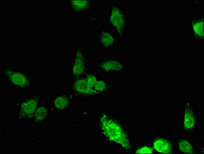
ApplicationsImmunoFluorescence, Western Blot
ReactivityHuman
TargetING5
- SizePrice

Product group Antibodies
Anti-ING5 Antibody Picoband(r)A04974-3-CARRIER-FREE
ApplicationsFlow Cytometry, ImmunoFluorescence, Western Blot, ELISA, ImmunoCytoChemistry
ReactivityHuman, Monkey
TargetING5
- SizePrice
Product group Antibodies
Anti-ING5 AntibodyA31978
ApplicationsImmunoFluorescence, Western Blot, ImmunoHistoChemistry
ReactivityHuman, Mouse
- SizePrice

Product group Antibodies
Anti-ING5 AntibodyHPA042685
ApplicationsImmunoHistoChemistry
ReactivityHuman
TargetING5
- SizePrice
Product group Antibodies

ING5 AntibodyLS-C346332
ApplicationsImmunoFluorescence, Western Blot, ImmunoHistoChemistry
ReactivityHuman
TargetING5
- SizePrice
Product group Antibodies
ING5 AntibodyCSB-PA820185LA01HU
ApplicationsImmunoFluorescence, ELISA
ReactivityHuman
TargetING5
- SizePrice
![ING5 antibody [N2C1], Internal detects ING5 protein by Western blot analysis. A. 30 μg Jurkat whole cell lysate/extract 12 % SDS-PAGE ING5 antibody [N2C1], Internal (GTX102482) dilution: 1:1000](https://www.genetex.com/upload/website/prouct_img/normal/GTX102482/GTX102482_39617_WB_w_23060100_570.webp)
Product group Antibodies
ING5 antibody [N2C1], InternalGTX102482
ApplicationsWestern Blot
ReactivityHuman
TargetING5
- SizePrice
Product group Antibodies
ING5 antibodyGTX33266
ApplicationsImmunoFluorescence, Western Blot, ImmunoCytoChemistry
ReactivityHuman
TargetING5
- SizePrice