

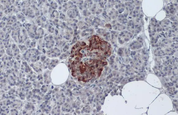
Insulin antibody detects Insulin protein at cytoplasm by immunohistochemical analysis. Sample: Paraffin-embedded human pancreas. Insulin stained by Insulin antibody (GTX27842) diluted at 1:50. Antigen Retrieval: Citrate buffer, pH 6.0, 15 min
 diluted at 1:50. Antigen Retrieval: Citrate buffer, pH 6.0, 15 min")
![Insulin antibody detects Insulin protein by immunohistochemical analysis. Sample: Frozen-sectioned mouse pancreas. Green: Insulin stained by Insulin antibody (GTX27842) diluted at 1:100. Red: C-Peptide antibody [HL1159] (GTX636463) diluted at 1:500. Blue: Fluoroshield with DAPI (GTX30920).](https://www.genetex.com/upload/website/prouct_img/normal/GTX27842/GTX27842_822500494_20250627_IHC-Fr_M_25070323_260.webp "Insulin antibody detects Insulin protein by immunohistochemical analysis. Sample: Frozen-sectioned mouse pancreas. Green: Insulin stained by Insulin antibody (GTX27842) diluted at 1:100. Red: C-Peptide antibody [HL1159] (GTX636463) diluted at 1:500. Blue: Fluoroshield with DAPI (GTX30920).")
Insulin antibody detects Insulin protein at cytoplasm by immunohistochemical analysis. Sample: Paraffin-embedded human pancreas. Insulin stained by Insulin antibody (GTX27842) diluted at 1:50. Antigen Retrieval: Citrate buffer, pH 6.0, 15 min
Insulin antibody
GTX27842
ApplicationsImmunoFluorescence, ImmunoCytoChemistry, ImmunoHistoChemistry, ImmunoHistoChemistry Frozen, ImmunoHistoChemistry Paraffin
Product group Antibodies
ReactivityHuman, Mouse
TargetINS
Overview
- SupplierGeneTex
- Product NameInsulin antibody
- Delivery Days Customer9
- Application Supplier NoteFor IHC/ICC: Use at a dilution of 1:50-1:100. Prolonged fixation in buffered formalin can destroy the epitope. Optimal dilutions/concentrations should be determined by the end user.
- ApplicationsImmunoFluorescence, ImmunoCytoChemistry, ImmunoHistoChemistry, ImmunoHistoChemistry Frozen, ImmunoHistoChemistry Paraffin
- CertificationResearch Use Only
- ClonalityPolyclonal
- Concentration0.2 mg/ml
- ConjugateUnconjugated
- Gene ID3630
- Target nameINS
- Target descriptioninsulin
- Target synonymsIDDM, IDDM1, IDDM2, ILPR, IRDN, MODY10, PNDM4, insulin, preproinsulin, proinsulin
- HostGuinea Pig
- IsotypeIgG
- Protein IDP01308
- Protein NameInsulin
- Scientific DescriptionAfter removal of the precursor signal peptide, proinsulin is post-translationally cleaved into three peptides: the B chain and A chain peptides, which are covalently linked via two disulfide bonds to form insulin, and C-peptide. Binding of insulin to the insulin receptor (INSR) stimulates glucose uptake. A multitude of mutant alleles with phenotypic effects have been identified. There is a read-through gene, INS-IGF2, which overlaps with this gene at the 5 region and with the IGF2 gene at the 3 region. Alternative splicing results in multiple transcript variants. [provided by RefSeq, Jun 2010]
- ReactivityHuman, Mouse
- Storage Instruction2°C to 8°C
- UNSPSC12352203
References
- Katz LS, Brill G, Wang P, et al. Transcriptional activation of the Myc gene by glucose in β-cells requires a ChREBP-dependent 3-D chromatin interaction between the Myc and Pvt1 genes. Mol Metab. 2024,79:101848. doi: 10.1016/j.molmet.2023.101848Read this paper
- Daian LM, Tanko G, Vacaru AM, et al. Modulation of Unfolded Protein Response Restores Survival and Function of β-Cells Exposed to the Endocrine Disruptor Bisphenol A. Int J Mol Sci. 2023,24(3). doi: 10.3390/ijms24032023Read this paper
- Morales-Reyes I, Atwater I, Esparza-Aguilar M, et al. Impact of biotin supplemented diet on mouse pancreatic islet β-cell mass expansion and glucose induced electrical activity. Islets. 2022,14(1):149-163. doi: 10.1080/19382014.2022.2091886Read this paper
- Lee SJ, Kim HJ, Byun NR, et al. Donor-Specific Regulatory T Cell-Mediated Immune Tolerance in an Intrahepatic Murine Allogeneic Islet Transplantation Model with Short-Term Anti-CD154 mAb Single Treatment. Cell Transplant. 2020,29:963689720913876. doi: 10.1177/0963689720913876Read this paper
- Chien HJ, Chiang TC, Peng SJ, et al. Human pancreatic afferent and efferent nerves: mapping and 3-D illustration of exocrine, endocrine, and adipose innervation. Am J Physiol Gastrointest Liver Physiol. 2019,317(5):G694-G706. doi: 10.1152/ajpgi.00116.2019Read this paper
- Nteeba J, Kubota K, Wang W, et al. Pancreatic prolactin receptor signaling regulates maternal glucose homeostasis. J Endocrinol. 2019,241(1):71-83. doi: 10.1530/JOE-18-0518Read this paper
- Tang SC, Shen CN, Lin PY, et al. Pancreatic neuro-insular network in young mice revealed by 3D panoramic histology. Diabetologia. 2018,61(1):158-167. doi: 10.1007/s00125-017-4408-yRead this paper
- Tang SC, Baeyens L, Shen CN, et al. Human pancreatic neuro-insular network in health and fatty infiltration. Diabetologia. 2018,61(1):168-181. doi: 10.1007/s00125-017-4409-xRead this paper
- Yamashita-Sugahara Y, Matsumoto M, Ohtaka M, et al. An inhibitor of fibroblast growth factor receptor-1 (FGFR1) promotes late-stage terminal differentiation from NGN3+ pancreatic endocrine progenitors. Sci Rep. 2016,6:35908. doi: 10.1038/srep35908Read this paper
- Lin PY, Peng SJ, Shen CN, et al. PanIN-associated pericyte, glial, and islet remodeling in mice revealed by 3D pancreatic duct lesion histology. Am J Physiol Gastrointest Liver Physiol. 2016,311(3):G412-22. doi: 10.1152/ajpgi.00071.2016Read this paper
Datasheet
Related products
Product group Antibodies

Anti-Insulin [HB125 (mAb1, AE9D6, Ab 125)]Ab03131-1.1
ApplicationsImmunoFluorescence, ELISA, ImmunoHistoChemistry
ReactivityHuman, Porcine, Rodent
TargetINS
- SizePrice
Product group Antibodies
Anti-Insulin Antibody130-11014
ApplicationsELISA
ReactivityHuman
TargetINS
- SizePrice

Product group Antibodies
References
ApplicationsImmunoFluorescence, ImmunoCytoChemistry, ImmunoHistoChemistry
ReactivityHuman, Mouse, Rat
TargetINS
- SizePrice

Product group Antibodies
ApplicationsELISA
ReactivityHuman
TargetINS
- SizePrice
Product group Antibodies
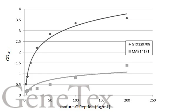
C-Peptide antibodyGTX129708
ApplicationsELISA, ImmunoHistoChemistry, ImmunoHistoChemistry Paraffin
ReactivityHuman, Mouse, Rat
TargetINS
- SizePrice
![Mouse tissue extract (50 μg) was separated by 15% SDS-PAGE, and the membrane was blotted with Insulin antibody [GT1229] (GTX02826) diluted at 1:1000. The HRP-conjugated anti-rabbit IgG antibody (GTX213110-01) was used to detect the primary antibody.](https://www.genetex.com/upload/website/prouct_img/normal/GTX02826/GTX02826_4000000209_20210122_WB_M_pancreas_w_23053123_391.webp)
Product group Antibodies
Insulin antibody [GT1229]GTX02826
ApplicationsWestern Blot, ImmunoHistoChemistry, ImmunoHistoChemistry Paraffin
ReactivityHuman, Mouse, Rat
TargetINS
- SizePrice
Product group Antibodies
C-Peptide antibody [GT1455]GTX631941
ApplicationsELISA
ReactivityHuman
TargetINS
- SizePrice
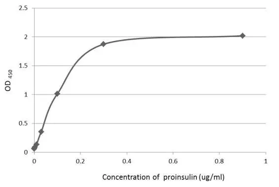
![An ELISA plate is coated with 50 μL of a Proinsulin recombinant protein at concentration ranged from 0.2 μg/mL to 1.6 μg/mL. The coated protein is detected with anti-C-peptide antibody [GT1489] (GTX631942) at 500 ng/mL.](https://www.genetex.com/upload/website/prouct_img/normal/GTX631942/GTX631942_41848_20150206_ELISA_w_23061202_525.webp)
Product group Antibodies
C-Peptide antibody [GT1489]GTX631942
ApplicationsELISA
ReactivityHuman
TargetINS
- SizePrice
![An ELISA plate is coated with 50 μL of a Proinsulin recombinant protein at concentration ranged from 0.2 μg/mL to 1.6 μg/mL. The coated protein is detected with anti-C-peptide antibody [GT1519] (GTX631943) at 500 ng/mL.](https://www.genetex.com/upload/website/prouct_img/normal/GTX631943/GTX631943_41848_20150206_ELISA_w_23061202_907.webp)
Product group Antibodies
C-Peptide antibody [GT1519]GTX631943
ApplicationsELISA
ReactivityHuman
TargetINS
- SizePrice