

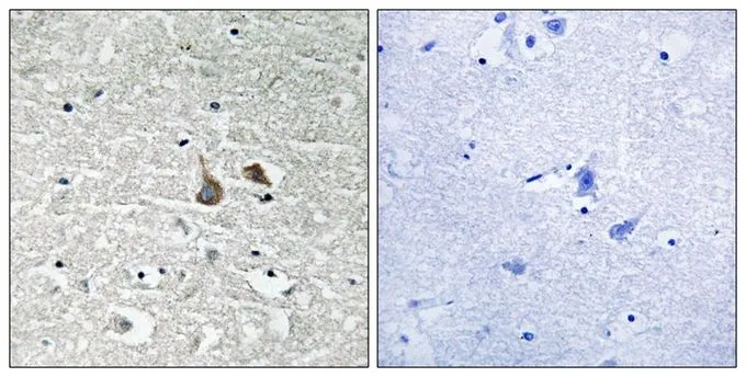
IHC-P analysis of human brain tissue using GTX55332 IRAK1 (phospho Ser376) antibody. Left : Primary antibody Right : Primary antibody pre-incubated with the antigen specific peptide
IHC-P analysis of human brain tissue using GTX55332 IRAK1 (phospho Ser376) antibody. Left : Primary antibody Right : Primary antibody pre-incubated with the antigen specific peptide
IRAK1 (phospho Ser376) antibody
GTX55332
ApplicationsWestern Blot, ImmunoHistoChemistry, ImmunoHistoChemistry Paraffin
Product group Antibodies
TargetIRAK1
Overview
- SupplierGeneTex
- Product NameIRAK1 (phospho Ser376) antibody
- Delivery Days Customer9
- Application Supplier NoteIHC-P: 1:50-1:100. *Optimal dilutions/concentrations should be determined by the researcher.Not tested in other applications.
- ApplicationsWestern Blot, ImmunoHistoChemistry, ImmunoHistoChemistry Paraffin
- CertificationResearch Use Only
- ClonalityPolyclonal
- Concentration1 mg/ml
- ConjugateUnconjugated
- Gene ID3654
- Target nameIRAK1
- Target descriptioninterleukin 1 receptor associated kinase 1
- Target synonymsinterleukin-1 receptor-associated kinase 1; IRAK; IRAK-1; pelle; Pelle homolog
- HostRabbit
- IsotypeIgG
- Scientific DescriptionThis gene encodes the interleukin-1 receptor-associated kinase 1, one of two putative serine/threonine kinases that become associated with the interleukin-1 receptor (IL1R) upon stimulation. This gene is partially responsible for IL1-induced upregulation of the transcription factor NF-kappa B. Alternatively spliced transcript variants encoding different isoforms have been found for this gene. [provided by RefSeq, Jul 2008]
- Storage Instruction-20°C or -80°C,2°C to 8°C
- UNSPSC12352203
References
- An IRAK1-PIN1 signalling axis drives intrinsic tumour resistance to radiation therapy. Liu PH et al., 2019 Feb, Nat Cell BiolRead more
Related products
Product group Antibodies
ApplicationsImmunoPrecipitation, Western Blot, ImmunoCytoChemistry, ImmunoHistoChemistry
TargetIRAK1
- SizePrice

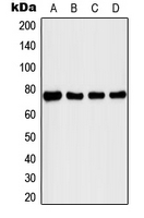
![WB analysis of mouse liver tissue lysate using GTX17299 IRAK1 antibody [8F1A7]. Working concentration : (A) 1 and (B) 2 microg/ml](https://www.genetex.com/upload/website/prouct_img/normal/GTX17299/GTX17299_2437_WB_20180221_w_23060620_282.webp)
Product group Antibodies
IRAK1 antibody [8F1A7]GTX17299
ApplicationsWestern Blot, ELISA, ImmunoHistoChemistry, ImmunoHistoChemistry Paraffin
TargetIRAK1
- SizePrice
Product group Antibodies
IRAK1 antibodyGTX20238
ApplicationsImmunoPrecipitation, Western Blot
TargetIRAK1
- SizePrice
Product group Antibodies
References
IRAK1 antibodyGTX31253
ApplicationsImmunoFluorescence, ImmunoPrecipitation, Western Blot, ELISA, ImmunoCytoChemistry
TargetIRAK1
- SizePrice

Product group Antibodies
Anti-IRAK1 AntibodyHPA054476
ApplicationsImmunoCytoChemistry, ImmunoHistoChemistry
TargetIRAK1
- SizePrice
Product group Antibodies
IRAK1 (phospho Thr209) antibodyGTX55098
ApplicationsWestern Blot
TargetIRAK1
- SizePrice
Product group Antibodies
IRAK1 AntibodyCSB-PA003061
ApplicationsImmunoFluorescence, Western Blot, ELISA
TargetIRAK1
- SizePrice