

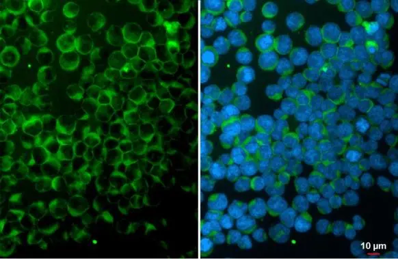
ITK antibody [N2C1], Internal detects ITK protein at cytoplasm by immunofluorescent analysis. Sample: Jurkat cells were fixed in 4% paraformaldehyde at RT for 15 min. Green: ITK stained by ITK antibody [N2C1], Internal (GTX113217) diluted at 1:500. Blue: Fluoroshield with DAPI (GTX30920).
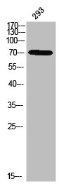
![Various whole cell extracts (30 μg) were separated by 7.5% SDS-PAGE, and the membrane was blotted with ITK antibody [N2C1], Internal (GTX113217) diluted at 1:1000. The HRP-conjugated anti-rabbit IgG antibody (GTX213110-01) was used to detect the primary antibody.](https://www.genetex.com/upload/website/prouct_img/normal/GTX113217/GTX113217_44203_20210122_WB_24010223_396.webp "Various whole cell extracts (30 μg) were separated by 7.5% SDS-PAGE, and the membrane was blotted with ITK antibody [N2C1], Internal (GTX113217) diluted at 1:1000. The HRP-conjugated anti-rabbit IgG antibody (GTX213110-01) was used to detect the primary antibody.")
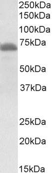
![Various whole cell extracts (30 μg) were separated by 7.5% SDS-PAGE, and the membrane was blotted with ITK antibody [N2C1], Internal (GTX113217) diluted at 1:1000. The HRP-conjugated anti-rabbit IgG antibody (GTX213110-01) was used to detect the primary antibody. Corresponding RNA expression data for the same cell lines are based on Human Protein Atlas program.](https://www.genetex.com/upload/website/prouct_img/normal/GTX113217/GTX113217_45497_20240809_WB_TPM_watermark_25112100_666.webp "Various whole cell extracts (30 μg) were separated by 7.5% SDS-PAGE, and the membrane was blotted with ITK antibody [N2C1], Internal (GTX113217) diluted at 1:1000. The HRP-conjugated anti-rabbit IgG antibody (GTX213110-01) was used to detect the primary antibody. Corresponding RNA expression data for the same cell lines are based on Human Protein Atlas program.")
ITK antibody [N2C1], Internal detects ITK protein at cytoplasm by immunofluorescent analysis. Sample: Jurkat cells were fixed in 4% paraformaldehyde at RT for 15 min. Green: ITK stained by ITK antibody [N2C1], Internal (GTX113217) diluted at 1:500. Blue: Fluoroshield with DAPI (GTX30920).
ITK antibody [N2C1], Internal
GTX113217
ApplicationsImmunoFluorescence, Western Blot, ImmunoCytoChemistry
Product group Antibodies
ReactivityHuman
TargetITK
Overview
- SupplierGeneTex
- Product NameITK antibody [N2C1], Internal
- Delivery Days Customer9
- Application Supplier NoteWB: 1:500-1:3000. ICC/IF: 1:100-1:1000. *Optimal dilutions/concentrations should be determined by the researcher.Not tested in other applications.
- ApplicationsImmunoFluorescence, Western Blot, ImmunoCytoChemistry
- CertificationResearch Use Only
- ClonalityPolyclonal
- Concentration0.25 mg/ml
- ConjugateUnconjugated
- Gene ID3702
- Target nameITK
- Target descriptionIL2 inducible T cell kinase
- Target synonymsEMT, LPFS1, LYK, PSCTK2, tyrosine-protein kinase ITK/TSK, IL-2-inducible T-cell kinase, T-cell-specific kinase, homolog of mouse T-cell itk/tsk, interleukin-2-inducible T-cell kinase, kinase EMT, tyrosine-protein kinase LYK
- HostRabbit
- IsotypeIgG
- Protein IDQ08881
- Protein NameTyrosine-protein kinase ITK/TSK
- Scientific DescriptionThis gene encodes an intracellular tyrosine kinase expressed in T-cells. The protein contains both SH2 and SH3 domains which are often found in intracellular kinases. It is thought to play a role in T-cell proliferation and differentiation. [provided by RefSeq]
- ReactivityHuman
- Storage Instruction-20°C or -80°C,2°C to 8°C
- UNSPSC41116161
Datasheet
Related products
Product group Antibodies
Phospho-ITK (Y512) AntibodyCSB-PA011277
ApplicationsWestern Blot, ELISA, ImmunoHistoChemistry
ReactivityHuman, Mouse
TargetITK
- SizePrice
Product group Antibodies
Itk Recombinant AntibodyCAC12298
ApplicationsELISA, ImmunoHistoChemistry
TargetITK
- SizePrice

Product group Antibodies
Anti-ITK Antibody Picoband(r)A01385-3-CARRIER-FREE
ApplicationsFlow Cytometry, Western Blot, ELISA, ImmunoHistoChemistry
ReactivityHuman
TargetITK
- SizePrice
Product group Antibodies
Anti-ITK/EMT AntibodyA84564
ApplicationsFlow Cytometry, ImmunoFluorescence, Western Blot, ELISA, ImmunoHistoChemistry
ReactivityHuman, Rat
- SizePrice
Product group Antibodies

Goat anti-ITKEB05117
ApplicationsImmunoFluorescence, Western Blot, ELISA
ReactivityHuman, Mouse
TargetITK
- SizePrice

Product group Antibodies
Anti-ITK AntibodyHPA043670
ApplicationsImmunoCytoChemistry
ReactivityHuman
TargetITK
- SizePrice
Product group Antibodies

ITK / EMT AntibodyLS-C405090
ApplicationsELISA, ImmunoHistoChemistry
ReactivityHuman, Mouse
TargetITK
- SizePrice
![WB analysis of truncated Trx-ITK recombinant protein using GTX83023 ITK antibody [5G12C4].](https://www.genetex.com/upload/website/prouct_img/normal/GTX83023/GTX83023_20170912_WB_w_23061322_874.webp)
Product group Antibodies
ITK antibody [5G12C4]GTX83023
ApplicationsWestern Blot, ELISA
ReactivityHuman
TargetITK
- SizePrice
![Whole cell extract (30 μg) was separated by 7.5% SDS-PAGE, and the membrane was blotted with ITK antibody [N3C3] (GTX113215) diluted at 1:500. The HRP-conjugated anti-rabbit IgG antibody (GTX213110-01) was used to detect the primary antibody.](https://www.genetex.com/upload/website/prouct_img/normal/GTX113215/GTX113215_40128_20170810_WB_w_23060501_333.webp)
Product group Antibodies
ITK antibody [N3C3]GTX113215
ApplicationsWestern Blot
ReactivityHuman
TargetITK
- SizePrice