

Immunohistochemical analysis of paraffin-embedded A549 xenograft, using Ketohexokinase(GTX109591) antibody at 1:500 dilution.
Antigen Retrieval: Citrate buffer, pH 6.0, 15 min
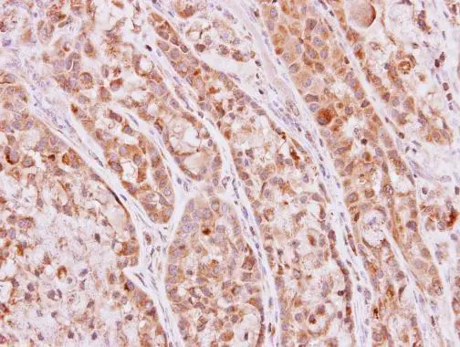
![Ketohexokinase antibody [N1C3] detects Ketohexokinase protein at cytoplasm by immunohistochemical analysis. Sample: Paraffin-embedded mouse liver. Ketohexokinase stained by Ketohexokinase antibody [N1C3] (GTX109591) diluted at 1:500. Antigen Retrieval: Citrate buffer, pH 6.0, 15 min](https://www.genetex.com/upload/website/prouct_img/normal/GTX109591/GTX109591_43867_20201211_IHC-P_M_w_23060500_305.webp "Ketohexokinase antibody [N1C3] detects Ketohexokinase protein at cytoplasm by immunohistochemical analysis. Sample: Paraffin-embedded mouse liver. Ketohexokinase stained by Ketohexokinase antibody [N1C3] (GTX109591) diluted at 1:500. Antigen Retrieval: Citrate buffer, pH 6.0, 15 min")
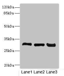
![Various whole cell extracts (30 μg) were separated by 12% SDS-PAGE, and the membrane was blotted with Ketohexokinase antibody [N1C3] (GTX109591) diluted at 1:1000. The HRP-conjugated anti-rabbit IgG antibody (GTX213110-01) was used to detect the primary antibody.](https://www.genetex.com/upload/website/prouct_img/normal/GTX109591/GTX109591_43867_20200227_WB_w_23060500_188.webp "Various whole cell extracts (30 μg) were separated by 12% SDS-PAGE, and the membrane was blotted with Ketohexokinase antibody [N1C3] (GTX109591) diluted at 1:1000. The HRP-conjugated anti-rabbit IgG antibody (GTX213110-01) was used to detect the primary antibody.")
![Various whole cell extracts (30 μg) were separated by 12% SDS-PAGE, and the membrane was blotted with Ketohexokinase antibody [N1C3] (GTX109591) diluted at 1:1000. The HRP-conjugated anti-rabbit IgG antibody (GTX213110-01) was used to detect the primary antibody, and the signal was developed with Trident ECL plus-Enhanced. Corresponding RNA expression data for the same cell lines are based on Human Protein Atlas program.](https://www.genetex.com/upload/website/prouct_img/normal/GTX109591/GTX109591_40030_20190920_WB_TPM_watermark_w_23060500_747.webp "Various whole cell extracts (30 μg) were separated by 12% SDS-PAGE, and the membrane was blotted with Ketohexokinase antibody [N1C3] (GTX109591) diluted at 1:1000. The HRP-conjugated anti-rabbit IgG antibody (GTX213110-01) was used to detect the primary antibody, and the signal was developed with Trident ECL plus-Enhanced. Corresponding RNA expression data for the same cell lines are based on Human Protein Atlas program.")
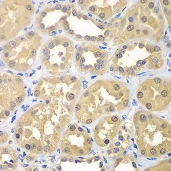
![Ketohexokinase antibody [N1C3] detects Ketohexokinase protein at cytoplasm and nucleus in mouse kidney by immunohistochemical analysis. Sample: Paraffin-embedded mouse kidney. Ketohexokinase antibody [N1C3] (GTX109591) diluted at 1:500.
Antigen Retrieval: Citrate buffer, pH 6.0, 15 min](https://www.genetex.com/upload/website/prouct_img/normal/GTX109591/GTX109591_40030_20150424_IHC_M_w_23060500_560.webp "Ketohexokinase antibody [N1C3] detects Ketohexokinase protein at cytoplasm and nucleus in mouse kidney by immunohistochemical analysis. Sample: Paraffin-embedded mouse kidney. Ketohexokinase antibody [N1C3] (GTX109591) diluted at 1:500.
Antigen Retrieval: Citrate buffer, pH 6.0, 15 min")
 A: mouse Liver 12% SDS PAGE GTX109591 diluted at 1:10000 The HRP-conjugated anti-rabbit IgG antibody (GTX213110-01) was used to detect the primary antibody.")


Immunohistochemical analysis of paraffin-embedded A549 xenograft, using Ketohexokinase(GTX109591) antibody at 1:500 dilution.
Antigen Retrieval: Citrate buffer, pH 6.0, 15 min
Ketohexokinase antibody [N1C3]
GTX109591
ApplicationsWestern Blot, ImmunoHistoChemistry, ImmunoHistoChemistry Paraffin
Product group Antibodies
ReactivityHuman, Mouse, Porcine, Rat
TargetKHK
Overview
- SupplierGeneTex
- Product NameKetohexokinase antibody [N1C3]
- Delivery Days Customer9
- Application Supplier NoteWB: 1:1000-1:10000. IHC-P: 1:100-1:1000. *Optimal dilutions/concentrations should be determined by the researcher.Not tested in other applications.
- ApplicationsWestern Blot, ImmunoHistoChemistry, ImmunoHistoChemistry Paraffin
- CertificationResearch Use Only
- ClonalityPolyclonal
- Concentration0.4 mg/ml
- ConjugateUnconjugated
- Gene ID3795
- Target nameKHK
- Target descriptionketohexokinase
- Target synonymsFRUCTU, ketohexokinase, fructokinase, hepatic fructokinase, testicular tissue protein Li 102
- HostRabbit
- IsotypeIgG
- Protein IDP50053
- Protein NameKetohexokinase
- Scientific DescriptionThis gene encodes ketohexokinase that catalyzes conversion of fructose to fructose-1-phosphate. The product of this gene is the first enzyme with a specialized pathway that catabolizes dietary fructose. Alternatively spliced transcript variants encoding different isoforms have been identified. [provided by RefSeq]
- ReactivityHuman, Mouse, Porcine, Rat
- Storage Instruction-20°C or -80°C,2°C to 8°C
- UNSPSC41116161
Datasheet
Related products
Product group Antibodies
Anti-KHK Antibody144-07440
ApplicationsWestern Blot, ImmunoHistoChemistry
ReactivityHuman, Mouse, Rat
TargetKHK
- SizePrice
Product group Antibodies
Anti-KHK AntibodyA32066
ApplicationsWestern Blot, ImmunoHistoChemistry
ReactivityMouse, Rat
- SizePrice
Product group Antibodies

KHK / Ketohexokinase AntibodyLS-C830227
ApplicationsWestern Blot, ELISA
ReactivityHuman
TargetKHK
- SizePrice
Product group Antibodies
ApplicationsImmunoFluorescence, Western Blot, ELISA, ImmunoCytoChemistry, ImmunoHistoChemistry, ImmunoHistoChemistry Frozen, ImmunoHistoChemistry Paraffin
ReactivityBovine, Equine, Human, Mouse, Porcine, Rabbit, Rat
TargetKHK
- SizePrice
Product group Antibodies
KHK AntibodyCSB-PA012157ESR1HU
ApplicationsImmunoPrecipitation, Western Blot, ELISA, ImmunoHistoChemistry
ReactivityHuman, Mouse, Rat
TargetKHK
- SizePrice
Product group Antibodies

Goat anti-KetohexokinaseEB12963
ApplicationsWestern Blot, ELISA, ImmunoHistoChemistry
ReactivityBovine, Canine, Human, Mouse, Porcine, Rat
TargetKHK
- SizePrice
Product group Antibodies
Khk Polyclonal AntibodyCAC10774
ApplicationsImmunoPrecipitation, Western Blot, ELISA, ImmunoHistoChemistry
ReactivityMouse, Rat
TargetKHK
- SizePrice
![IHC-P analysis of human colon adenocarcinoma tissue using GTX84277 Ketohexokinase antibody [3D1]. Antigen retrieval : Heat-induced epitope retrieval by 10mM citrate buffer, pH6.0, 100oC for 10min.](https://www.genetex.com/upload/website/prouct_img/normal/GTX84277/GTX84277_2517_IHC-P_w_23061420_156.webp)
Product group Antibodies
Ketohexokinase antibody [3D1]GTX84277
ApplicationsFlow Cytometry, ImmunoFluorescence, Western Blot, ImmunoCytoChemistry, ImmunoHistoChemistry, ImmunoHistoChemistry Paraffin
ReactivityHuman
TargetKHK
- SizePrice

Product group Antibodies
Anti-KHK-25ulHPA007040
ApplicationsWestern Blot, ImmunoHistoChemistry
ReactivityHuman, Mouse, Rat
- SizePrice
Product group Antibodies
Ketohexokinase antibodyGTX33283
ApplicationsWestern Blot, ImmunoHistoChemistry, ImmunoHistoChemistry Paraffin
ReactivityHuman, Mouse, Rat
TargetKHK
- SizePrice