

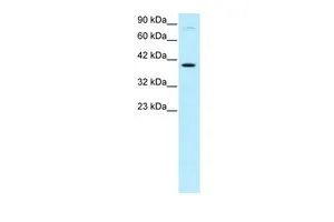
WB analysis of HepG2 cells using GTX77858 KIN antibody at 0.2-1μg/ml.


WB analysis of HepG2 cells using GTX77858 KIN antibody at 0.2-1μg/ml.
KIN antibody, Internal
GTX77858
ApplicationsWestern Blot, ImmunoHistoChemistry, ImmunoHistoChemistry Paraffin
Product group Antibodies
ReactivityHuman
TargetKIN
Overview
- SupplierGeneTex
- Product NameKIN antibody, Internal
- Delivery Days Customer9
- Application Supplier NoteWB: 0.2-2.5 ug/ml. IHC-P: 2-10 ug/ml. *Optimal dilutions/concentrations should be determined by the researcher.Not tested in other applications.
- ApplicationsWestern Blot, ImmunoHistoChemistry, ImmunoHistoChemistry Paraffin
- CertificationResearch Use Only
- ClonalityPolyclonal
- Concentration0.5-1 mg/ml
- ConjugateUnconjugated
- Gene ID22944
- Target nameKIN
- Target descriptionKin17 DNA and RNA binding protein
- Target synonymsBTCD, KIN17, Rts2, DNA/RNA-binding protein KIN17, KIN, antigenic determinant of recA protein homolog, binding to curved DNA
- HostRabbit
- IsotypeIgG
- Protein IDO60870
- Protein NameDNA/RNA-binding protein KIN17
- Scientific DescriptionThe protein encoded by this gene is a nuclear protein that forms intranuclear foci during proliferation and is redistributed in the nucleoplasm during the cell cycle. Short-wave ultraviolet light provokes the relocalization of the protein, suggesting its participation in the cellular response to DNA damage. Originally selected based on protein-binding with RecA antibodies, the mouse protein presents a limited similarity with a functional domain of the bacterial RecA protein, a characteristic shared by this human ortholog. Alternative splicing of this gene results in multiple transcript variants. [provided by RefSeq, Jan 2012]
- ReactivityHuman
- Storage Instruction-20°C or -80°C,2°C to 8°C
- UNSPSC41116161
Datasheet
Related products
Product group Antibodies
KIN AntibodyCSB-PA012351GA01HU
ApplicationsWestern Blot, ELISA
ReactivityHuman, Mouse, Rat
TargetKIN
- SizePrice
Product group Antibodies
Anti-KIN AntibodyA306061
ApplicationsWestern Blot
ReactivityHuman, Mouse, Rat
- SizePrice

Product group Antibodies
Anti-KIN AntibodyHPA038700
ApplicationsChIP Chromatin ImmunoPrecipitation, ImmunoCytoChemistry
ReactivityHuman
TargetKIN
- SizePrice
Product group Antibodies

KIN17 / KIN Antibody (aa48-64)LS-C313337
ApplicationsWestern Blot
ReactivityBovine, Chicken, Equine, Guinea Pig, Human, Mammals, Monkey, Mouse, Rabbit, Rat, Sheep, Xenopus
TargetKIN
- SizePrice

Product group Antibodies
Anti-KIN Antibody Picoband(r)PB9607-CARRIER-FREE
ApplicationsWestern Blot, ImmunoHistoChemistry
ReactivityHuman, Mouse, Rat
TargetKIN
- SizePrice