

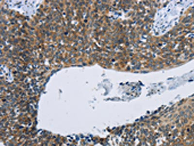
The image on the left is immunohistochemistry of paraffin-embedded Human cervical cancer tissue using CSB-PA244093(KPNB1 Antibody) at dilution 1/40, on the right is treated with synthetic peptide. (Original magnification: x200)
 at dilution 1/40, on the right is treated with synthetic peptide. (Original magnification: x200)")
The image on the left is immunohistochemistry of paraffin-embedded Human cervical cancer tissue using CSB-PA244093(KPNB1 Antibody) at dilution 1/40, on the right is treated with synthetic peptide. (Original magnification: x200)
KPNB1 Antibody
CSB-PA244093
ApplicationsELISA, ImmunoHistoChemistry
Product group Antibodies
ReactivityHuman, Mouse, Rat
TargetKPNB1
Overview
- SupplierCusabio
- Product NameKPNB1 Antibody
- Delivery Days Customer20
- ApplicationsELISA, ImmunoHistoChemistry
- CertificationResearch Use Only
- ClonalityPolyclonal
- ConjugateUnconjugated
- Gene ID3837
- Target nameKPNB1
- Target descriptionkaryopherin subunit beta 1
- Target synonymsIMB1, IPO1, IPOB, Impnb, NTF97, importin subunit beta-1, PTAC97, importin 1, importin 90, importin beta-1 subunit, karyopherin (importin) beta 1, nuclear factor p97, pore targeting complex 97 kDa subunit
- HostRabbit
- IsotypeIgG
- Protein IDQ14974
- Protein NameImportin subunit beta-1
- Scientific DescriptionNucleocytoplasmic transport, a signal- and energy-dependent process, takes place through nuclear pore complexes embedded in the nuclear envelope. The import of proteins containing a nuclear localization signal (NLS) requires the NLS import receptor, a heterodimer of importin alpha and beta subunits also known as karyopherins. Importin alpha binds the NLS-containing cargo in the cytoplasm and importin beta docks the complex at the cytoplasmic side of the nuclear pore complex. In the presence of nucleoside triphosphates and the small GTP binding protein Ran, the complex moves into the nuclear pore complex and the importin subunits dissociate. Importin alpha enters the nucleoplasm with its passenger protein and importin beta remains at the pore.
- ReactivityHuman, Mouse, Rat
- Storage Instruction-20°C or -80°C
- UNSPSC41116161
Related products
Product group Antibodies
Anti-KPNB1 AntibodyA16248
ApplicationsImmunoFluorescence, Western Blot, ImmunoCytoChemistry, ImmunoHistoChemistry
ReactivityHuman, Mouse, Rat
- SizePrice
Product group Antibodies
Anti-KPNB1 Antibody144-08610
ApplicationsWestern Blot, ImmunoHistoChemistry
ReactivityHuman, Mouse, Rat
TargetKPNB1
- SizePrice

Product group Antibodies
Anti-KPNB1 Antibody Picoband(r)A01851-2-CARRIER-FREE
ApplicationsFlow Cytometry, ImmunoFluorescence, Western Blot, ELISA, ImmunoCytoChemistry
ReactivityHuman, Mouse, Rat
TargetKPNB1
- SizePrice
Product group Antibodies
NTF97 Polyclonal AntibodyBS-3695R
ApplicationsImmunoFluorescence, Western Blot, ELISA, ImmunoCytoChemistry, ImmunoHistoChemistry, ImmunoHistoChemistry Frozen, ImmunoHistoChemistry Paraffin
ReactivityBovine, Chicken, Equine, Human, Mouse, Porcine, Rat, Sheep
TargetKPNB1
- SizePrice
Product group Antibodies

ApplicationsFlow Cytometry, ImmunoFluorescence, Western Blot, ELISA, ImmunoHistoChemistry
ReactivityBovine, Canine, Human, Mouse, Rat
TargetKPNB1
- SizePrice
Product group Antibodies
KPNB1 Polyclonal AntibodyCAC15768
ApplicationsWestern Blot, ELISA, ImmunoHistoChemistry
TargetKPNB1
- SizePrice
Product group Antibodies

KPNB1 / Importin Beta AntibodyLS-C403249
ApplicationsELISA, ImmunoHistoChemistry
ReactivityHuman, Mouse, Rat
TargetKPNB1
- SizePrice
Product group Antibodies
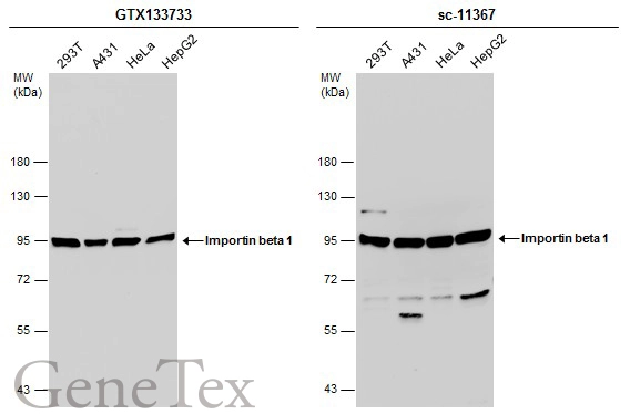
Importin beta 1 antibodyGTX133733
ApplicationsWestern Blot, ImmunoHistoChemistry, ImmunoHistoChemistry Paraffin
ReactivityHuman, Mouse
TargetKPNB1
- SizePrice

Product group Antibodies
Anti-KPNB1 AntibodyHPA050302
ApplicationsImmunoCytoChemistry
ReactivityHuman
TargetKPNB1
- SizePrice
Product group Antibodies
Anti-KPNB1 AntibodyCAB8610
ApplicationsImmunoFluorescence, Western Blot, ELISA, ImmunoCytoChemistry, ImmunoHistoChemistry, ImmunoHistoChemistry Paraffin
ReactivityHuman
TargetKPNB1
- SizePrice