

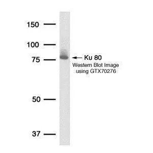
Detection of human Ku80 protein in HeLa whole cell lysate using GeneTex Ku80 149.8 monoclonal antibody GTX70276.
 A: 293T 20ug B: 293T 10ug C: 293T 5ug 7.5% SDS PAGE GTX70276 diluted at 1:10000")
![Various whole cell extracts (30 μg) were separated by 7.5% SDS-PAGE, and the membrane was blotted with Ku80 antibody [149.8] (GTX70276) diluted at 1:1000. The HRP-conjugated anti-mouse IgG antibody (GTX213111-01) was used to detect the primary antibody.](https://www.genetex.com/upload/website/prouct_img/normal/GTX70276/GTX70276_40980_20211105_WB_w_23061221_172.webp "Various whole cell extracts (30 μg) were separated by 7.5% SDS-PAGE, and the membrane was blotted with Ku80 antibody [149.8] (GTX70276) diluted at 1:1000. The HRP-conjugated anti-mouse IgG antibody (GTX213111-01) was used to detect the primary antibody.")
 A: Jurkat B: Raji C: 293T D: A431 E: HeLa F: HepG2 G: H1299 H: HCT116 I: MCF-7 J: NT2D1 K: PC-3 L: U87-MG 7.5% SDS PAGE GTX70276 diluted at 1:10000")
![Ku80 antibody [149.8] detects Ku80 protein at nucleus by immunofluorescent analysis. Sample: HeLa cells were fixed in 4% paraformaldehyde at RT for 15 min. Green: Ku80 protein stained by Ku80 antibody [149.8] (GTX70276) diluted at 1:500. Blue: Hoechst 33343 staining.](https://www.genetex.com/upload/website/prouct_img/normal/GTX70276/GTX70276_40980_IFA_w_23061221_685.webp "Ku80 antibody [149.8] detects Ku80 protein at nucleus by immunofluorescent analysis. Sample: HeLa cells were fixed in 4% paraformaldehyde at RT for 15 min. Green: Ku80 protein stained by Ku80 antibody [149.8] (GTX70276) diluted at 1:500. Blue: Hoechst 33343 staining.")


Detection of human Ku80 protein in HeLa whole cell lysate using GeneTex Ku80 149.8 monoclonal antibody GTX70276.
Ku80 antibody [149.8]
GTX70276
ApplicationsImmunoFluorescence, Western Blot, ImmunoCytoChemistry
Product group Antibodies
ReactivityHuman, Monkey
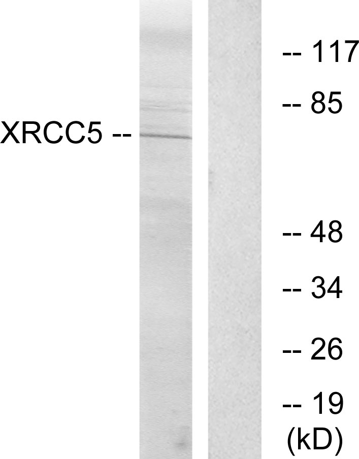
TargetXRCC5
Overview
- SupplierGeneTex
- Product NameKu80 antibody [149.8]
- Delivery Days Customer9
- Application Supplier NoteWB: 1:1000-1:10000. ICC/IF: 1:100-1:1000. *Optimal dilutions/concentrations should be determined by the researcher.Not tested in other applications.
- ApplicationsImmunoFluorescence, Western Blot, ImmunoCytoChemistry
- CertificationResearch Use Only
- ClonalityMonoclonal
- Clone ID149.8
- Concentration1 mg/ml
- ConjugateUnconjugated
- Gene ID7520
- Target nameXRCC5
- Target descriptionX-ray repair cross complementing 5
- Target synonymsKARP-1, KARP1, KU80, KUB2, Ku86, NFIV, X-ray repair cross-complementing protein 5, 86 kDa subunit of Ku antigen, ATP-dependent DNA helicase 2 subunit 2, ATP-dependent DNA helicase II 80 kDa subunit, CTC box-binding factor 85 kDa subunit, CTC85, CTCBF, DNA repair protein XRCC5, Ku autoantigen, 80kDa, Ku86 autoantigen related protein 1, TLAA, X-ray repair complementing defective repair in Chinese hamster cells 5 (double-strand-break rejoining), lupus Ku autoantigen protein p86, nuclear factor IV, thyroid-lupus autoantigen
- HostMouse
- IsotypeIgG1
- Protein IDP13010
- Protein NameX-ray repair cross-complementing protein 5
- Scientific DescriptionThe protein encoded by this gene is the 80-kilodalton subunit of the Ku heterodimer protein which is also known as ATP-dependant DNA helicase II or DNA repair protein XRCC5. Ku is the DNA-binding component of the DNA-dependent protein kinase, and it functions together with the DNA ligase IV-XRCC4 complex in the repair of DNA double-strand break by non-homologous end joining and the completion of V(D)J recombination events. This gene functionally complements Chinese hamster xrs-6, a mutant defective in DNA double-strand break repair and in ability to undergo V(D)J recombination. A rare microsatellite polymorphism in this gene is associated with cancer in patients of varying radiosensitivity. [provided by RefSeq, Jul 2008]
- ReactivityHuman, Monkey
- Storage Instruction-20°C or -80°C,2°C to 8°C
- UNSPSC41116161
Datasheet
Related products
Product group Antibodies

Ku80 AntibodyABX013131
ApplicationsImmunoFluorescence, Western Blot, ELISA, ImmunoCytoChemistry, ImmunoHistoChemistry
- SizePrice
Product group Antibodies
Anti-XRCC5 AntibodyA100714
ApplicationsWestern Blot, ELISA
ReactivityHuman
- SizePrice
Product group Antibodies
Anti-XRCC5 Antibody144-05862
ApplicationsImmunoFluorescence, ImmunoPrecipitation, Western Blot, ImmunoHistoChemistry
ReactivityHuman
TargetXRCC5
- SizePrice
Product group Antibodies
Ku80 Recombinant AntibodyBSM-52512R
ApplicationsImmunoFluorescence, Western Blot, ImmunoCytoChemistry, ImmunoHistoChemistry, ImmunoHistoChemistry Frozen, ImmunoHistoChemistry Paraffin
ReactivityHuman
TargetXRCC5
- SizePrice
Product group Antibodies
XRCC5 AntibodyCSB-PA003114
ApplicationsImmunoFluorescence, Western Blot, ELISA, ImmunoHistoChemistry
ReactivityHuman, Monkey
TargetXRCC5
- SizePrice
Product group Antibodies

Goat anti-Ku80 / XRCC5EB07209
ApplicationsWestern Blot, ELISA, ImmunoHistoChemistry
ReactivityHuman
TargetXRCC5
- SizePrice
Product group Antibodies
XRCC5 Polyclonal AntibodyCAC14001
ApplicationsWestern Blot, ELISA, ImmunoHistoChemistry
TargetXRCC5
- SizePrice
![IHC-P analysis of human mucosa from appendix tissue using GTX04480 Ku80 antibody [MSVA-880M] HistoMAX?. Appendix mucosa showing a strong nuclear Ku80 staining in all cells.](https://www.genetex.com/upload/website/prouct_img/normal/GTX04480/GTX04480_20230728_IHC-P_129_23072722_291.webp)
Product group Antibodies
ApplicationsImmunoHistoChemistry, ImmunoHistoChemistry Paraffin
ReactivityHuman
TargetXRCC5
- SizePrice
Product group Antibodies

XRCC5 / Ku80 Antibody (C-Terminus)LS-C368653
ApplicationsImmunoFluorescence, Western Blot, ImmunoCytoChemistry, ImmunoHistoChemistry, ImmunoHistoChemistry Paraffin
ReactivityHuman, Monkey
TargetXRCC5
- SizePrice
Product group Antibodies
Ku80 antibodyGTX70485
ApplicationsWestern Blot, ELISA
ReactivityHuman, Mouse
- SizePrice