

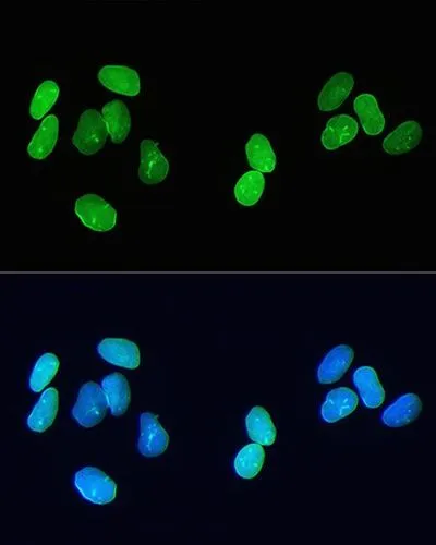
ICC/IF analysis of U2OS cells using GTX00774 Lamin A + C antibody [GT1137]. Green: Primary antibody Blue: DAPI Dlution : 1:200
![Various whole cell extracts (30 μg) were separated by 10% SDS-PAGE, and the membrane was blotted with Lamin A + C antibody [GT1137] (GTX00774) diluted at 1:500. The HRP-conjugated anti-rabbit IgG antibody (GTX213110-01) was used to detect the primary antibody.](https://www.genetex.com/upload/website/prouct_img/normal/GTX00774/GTX00774_3560448007_20200313_WB_w_23053121_414.webp "Various whole cell extracts (30 μg) were separated by 10% SDS-PAGE, and the membrane was blotted with Lamin A + C antibody [GT1137] (GTX00774) diluted at 1:500. The HRP-conjugated anti-rabbit IgG antibody (GTX213110-01) was used to detect the primary antibody.")
![WB analysis of various samples using GTX00774 Lamin A + C antibody [GT1137]. Dilution : 1:50000 Loading : 25μg](https://www.genetex.com/upload/website/prouct_img/normal/GTX00774/GTX00774_20200327_WB_2_w_23053121_343.webp "WB analysis of various samples using GTX00774 Lamin A + C antibody [GT1137]. Dilution : 1:50000 Loading : 25μg")
![WB analysis of NIH-3T3 extracts using GTX00774 Lamin A + C antibody [GT1137]. WCE : whole cell lysate CE : cytoplasmic extracts NE : nuclear extracts Dilution : 1:10000 Loading : 25μg](https://www.genetex.com/upload/website/prouct_img/normal/GTX00774/GTX00774_20200327_WB_4_w_23053121_568.webp "WB analysis of NIH-3T3 extracts using GTX00774 Lamin A + C antibody [GT1137]. WCE : whole cell lysate CE : cytoplasmic extracts NE : nuclear extracts Dilution : 1:10000 Loading : 25μg")
![ICC/IF analysis of HeLa cells using GTX00774 Lamin A + C antibody [GT1137]. Red: Primary antibody Blue: DAPI Detection system: confocol Dlution : 1:100](https://www.genetex.com/upload/website/prouct_img/normal/GTX00774/GTX00774_20191101_AP_002_20_w_23053121_867.webp "ICC/IF analysis of HeLa cells using GTX00774 Lamin A + C antibody [GT1137]. Red: Primary antibody Blue: DAPI Detection system: confocol Dlution : 1:100")
![WB analysis of HeLa whole cell lysate using GTX00774 Lamin A + C antibody [GT1137]. Dilution :1:10000-1:2560000 Loading : 25μg](https://www.genetex.com/upload/website/prouct_img/normal/GTX00774/GTX00774_20200327_WB_1_w_23053121_865.webp "WB analysis of HeLa whole cell lysate using GTX00774 Lamin A + C antibody [GT1137]. Dilution :1:10000-1:2560000 Loading : 25μg")
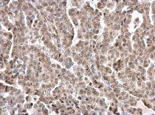
![IHC-P analysis of hmuan breast cancer tissue section using GTX00774 Lamin A + C antibody [GT1137]. Dlution : 1:200](https://www.genetex.com/upload/website/prouct_img/normal/GTX00774/GTX00774_20191101_AP_003_15_w_23053121_217.webp "IHC-P analysis of hmuan breast cancer tissue section using GTX00774 Lamin A + C antibody [GT1137]. Dlution : 1:200")
![IP analysis of K562 whole cell lysate using GTX00774 Lamin A + C antibody [GT1137]. Total protein amount : 200μg IP antibody : 3μg Dilution : 1:20000](https://www.genetex.com/upload/website/prouct_img/normal/GTX00774/GTX00774_20200327_IP_7_w_23053121_725.webp "IP analysis of K562 whole cell lysate using GTX00774 Lamin A + C antibody [GT1137]. Total protein amount : 200μg IP antibody : 3μg Dilution : 1:20000")
![ICC/IF analysis of U2OS cells using GTX00774 Lamin A + C antibody [GT1137]. Pink: Primary antibody Blue: DAPI Dlution : 1:100-1:1600](https://www.genetex.com/upload/website/prouct_img/normal/GTX00774/GTX00774_20191101_AP_002_21_w_23053121_755.webp "ICC/IF analysis of U2OS cells using GTX00774 Lamin A + C antibody [GT1137]. Pink: Primary antibody Blue: DAPI Dlution : 1:100-1:1600")
![ICC/IF analysis of L929 cells using GTX00774 Lamin A + C antibody [GT1137]. Green: Primary antibody Blue: DAPI The image was taken by Confocal microscopy. Dlution : 1:200](https://www.genetex.com/upload/website/prouct_img/normal/GTX00774/GTX00774_20191101_AP_002_19_w_23053121_348.webp "ICC/IF analysis of L929 cells using GTX00774 Lamin A + C antibody [GT1137]. Green: Primary antibody Blue: DAPI The image was taken by Confocal microscopy. Dlution : 1:200")
ICC/IF analysis of U2OS cells using GTX00774 Lamin A + C antibody [GT1137]. Green: Primary antibody Blue: DAPI Dlution : 1:200
Lamin A + C antibody [GT1137]
GTX00774
ApplicationsImmunoFluorescence, ImmunoPrecipitation, Western Blot, ImmunoCytoChemistry, ImmunoHistoChemistry, ImmunoHistoChemistry Paraffin
Product group Antibodies
ReactivityHuman, Mouse, Rat
TargetLMNA
Overview
- SupplierGeneTex
- Product NameLamin A + C antibody [GT1137]
- Delivery Days Customer9
- Application Supplier NoteWB: 1:50000 - 1:200000. ICC/IF: 1:50 - 1:1000. IHC-P: 1:50 - 1:1000. IP: 1:50 - 1:200. *Optimal dilutions/concentrations should be determined by the researcher.Not tested in other applications.
- ApplicationsImmunoFluorescence, ImmunoPrecipitation, Western Blot, ImmunoCytoChemistry, ImmunoHistoChemistry, ImmunoHistoChemistry Paraffin
- CertificationResearch Use Only
- ClonalityMonoclonal
- Clone IDGT1137
- ConjugateUnconjugated
- Gene ID4000
- Target nameLMNA
- Target descriptionlamin A/C
- Target synonymsCDCD1, CDDC, CMD1A, CMT2B1, EMD2, FPL, FPLD, FPLD2, HGPS, IDC, LDP1, LFP, LGMD1B, LMN1, LMNC, LMNL1, MADA, PRO1, lamin, 70 kDa lamin, epididymis secretory sperm binding protein, lamin A/C-like 1, lamin C, mandibuloacral dysplasia type A, prelamin-A/C, progerin, renal carcinoma antigen NY-REN-32
- HostRabbit
- IsotypeIgG
- Protein IDP02545
- Protein NamePrelamin-A/C
- Scientific DescriptionThe nuclear lamina consists of a two-dimensional matrix of proteins located next to the inner nuclear membrane. The lamin family of proteins make up the matrix and are highly conserved in evolution. During mitosis, the lamina matrix is reversibly disassembled as the lamin proteins are phosphorylated. Lamin proteins are thought to be involved in nuclear stability, chromatin structure and gene expression. Vertebrate lamins consist of two types, A and B. Alternative splicing results in multiple transcript variants. Mutations in this gene lead to several diseases: Emery-Dreifuss muscular dystrophy, familial partial lipodystrophy, limb girdle muscular dystrophy, dilated cardiomyopathy, Charcot-Marie-Tooth disease, and Hutchinson-Gilford progeria syndrome. [provided by RefSeq, Apr 2012]
- ReactivityHuman, Mouse, Rat
- Storage Instruction-20°C or -80°C,2°C to 8°C
- UNSPSC12352203
Datasheet
Related products
Product group Antibodies

Anti-Lamin A [133A2]AB03358-3.0
ApplicationsFlow Cytometry, ImmunoFluorescence, Western Blot, ImmunoHistoChemistry
ReactivityBovine, Canine, Human, Mouse, Rat
TargetLMNA
- SizePrice
Product group Antibodies
Anti-LMNA Antibody144-00249
ApplicationsImmunoFluorescence, Western Blot, ImmunoHistoChemistry
ReactivityHuman, Mouse, Rat
TargetLMNA
- SizePrice
Product group Antibodies
References
Lamin A + C antibodyGTX101126
ApplicationsImmunoFluorescence, ImmunoPrecipitation, Western Blot, ImmunoCytoChemistry, ImmunoHistoChemistry, ImmunoHistoChemistry Paraffin
ReactivityHuman, Mouse
TargetLMNA
- SizePrice
Product group Antibodies
References
Lamin A + C antibodyGTX101127
ApplicationsImmunoFluorescence, ImmunoPrecipitation, Western Blot, ImmunoCytoChemistry, ImmunoHistoChemistry, ImmunoHistoChemistry Paraffin
ReactivityHuman, Mouse, Rat
TargetLMNA
- SizePrice
Product group Antibodies
References
Lamin A + C antibodyGTX111677
ApplicationsWestern Blot, ImmunoHistoChemistry, ImmunoHistoChemistry Paraffin
ReactivityHuman, Mouse
TargetLMNA
- SizePrice
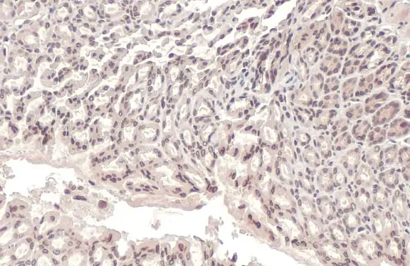
![IHC-P analysis of human striated muscle tissue using GTX60478 Lamin A antibody [4E7].](https://www.genetex.com/upload/website/prouct_img/normal/GTX60478/GTX60478_20170912_IHC-P_w_23061123_665.webp)
Product group Antibodies
Lamin A antibody [4E7]GTX60478
ApplicationsWestern Blot, ELISA, ImmunoHistoChemistry, ImmunoHistoChemistry Paraffin
ReactivityHuman, Mouse, Rat
TargetLMNA
- SizePrice
![Lamin A + C antibody [GT9712] detects Lamin A + C protein at nuclear envelope by immunofluorescent analysis. Sample: HeLa cells were fixed in ice-cold MeOH for 5 min. Green: Lamin A + C stained by Lamin A + C antibody [GT9712] (GTX629404) diluted at 1:500. Red: beta Tubulin, a cytoskeleton marker, stained by beta Tubulin antibody (GTX101279) diluted at 1:1000.](https://www.genetex.com/upload/website/prouct_img/normal/GTX629404/GTX629404_44748_20220902_ICC_IF_22101717_764.webp)
Product group Antibodies
Lamin A + C antibody [GT9712]GTX629404
ApplicationsImmunoFluorescence, ImmunoPrecipitation, Western Blot, ImmunoCytoChemistry
ReactivityHuman
TargetLMNA
- SizePrice
![ICC/IF analysis of Human fibroblast cells using GTX80813 Lamin A + C antibody [mab636]. Fixation : methanol Permeabilization : 0.1% Triton X-100 in TBS for 10 minutes Dilution : 1:200 for at least 1 hour at room temperature](https://www.genetex.com/upload/website/prouct_img/normal/GTX80813/GTX80813_910_ICC-IF_w_23061322_876.webp)
Product group Antibodies
Lamin A + C antibody [mab636]GTX80813
ApplicationsImmunoFluorescence, Western Blot, ImmunoCytoChemistry, ImmunoHistoChemistry, ImmunoHistoChemistry Frozen
ReactivityBovine, Canine, Human, Mouse, Porcine
TargetLMNA
- SizePrice
Product group Antibodies
ApplicationsWestern Blot, ImmunoHistoChemistry, ImmunoHistoChemistry Paraffin
ReactivityHuman
TargetLMNA
- SizePrice