

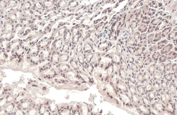
Lamin A + C antibody detects Lamin A + C protein at cytoplasm and nucleus by immunohistochemical analysis. Sample: Paraffin-embedded mouse stomach. Lamin A + C stained by Lamin A + C antibody (GTX101127) diluted at 1:100. Antigen Retrieval: Citrate buffer, pH 6.0, 15 min
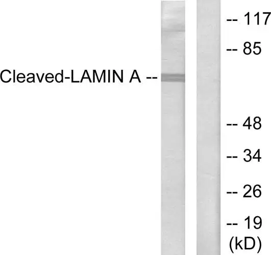
 and Lamin A + C knockout (KO) HeLa cell extracts (30 μg) were separated by 7.5% SDS-PAGE, and the membrane was blotted with Lamin A + C antibody (GTX101127) diluted at 1:1000. The HRP-conjugated anti-rabbit IgG antibody (GTX213110-01) was used to detect the primary antibody.")
![Lamin A + C antibody immunoprecipitates Lamin A + C protein in IP experiments. IP samples: HeLa whole cell extract A. 50 μg HeLa whole cell extract B. Control with 4 μg of preimmune Rabbit IgG C. Immunoprecipitation of Lamin A + C protein by 4 μg Lamin A + C antibody (GTX101127) 7.5 % SDS-PAGE The immunoprecipitated Lamin A + C protein was detected by Lamin A + C antibody (GTX101127) diluted at 1:500. [EasyBlot anti-rabbit IgG (GTX221666-01) was used as a secondary reagent]](https://www.genetex.com/upload/website/prouct_img/normal/GTX101127/GTX101127_40401_IP_w_23060100_555.webp "Lamin A + C antibody immunoprecipitates Lamin A + C protein in IP experiments. IP samples: HeLa whole cell extract A. 50 μg HeLa whole cell extract B. Control with 4 μg of preimmune Rabbit IgG C. Immunoprecipitation of Lamin A + C protein by 4 μg Lamin A + C antibody (GTX101127) 7.5 % SDS-PAGE The immunoprecipitated Lamin A + C protein was detected by Lamin A + C antibody (GTX101127) diluted at 1:500. [EasyBlot anti-rabbit IgG (GTX221666-01) was used as a secondary reagent]")
 antibody at 1:500 dilution.
Antigen Retrieval: Trilogy? (EDTA based, pH 8.0) buffer, 15min")
 A: NIH-3T3 7.5% SDS PAGE GTX101127 diluted at 1:1000 The HRP-conjugated anti-rabbit IgG antibody (GTX213110-01) was used to detect the primary antibody.")
 antibody at 1:500 dilution.
Antigen Retrieval: Trilogy? (EDTA based, pH 8.0) buffer, 15min")
 were separated by 7.5% SDS-PAGE, and the membrane was blotted with Lamin A + C antibody (GTX101127) diluted at 1:2000. The HRP-conjugated anti-rabbit IgG antibody (GTX213110-01) was used to detect the primary antibody. Corresponding RNA expression data for the same cell lines are based on Human Protein Atlas program.")
![Lamin A + C antibody detects Lamin A + C protein at nuclear envelope by immunofluorescent analysis. Sample: HeLa cells were fixed in ice-cold MeOH for 5 min. Green: Lamin A + C stained by Lamin A + C antibody (GTX101127) diluted at 1:1000. Red: alpha Tubulin, stained by alpha Tubulin antibody [GT114] (GTX628802) diluted at 1:500. Blue: Fluoroshield with DAPI (GTX30920).](https://www.genetex.com/upload/website/prouct_img/normal/GTX101127/GTX101127_43460_20190410_ICC_IF_w_23060100_490.webp "Lamin A + C antibody detects Lamin A + C protein at nuclear envelope by immunofluorescent analysis. Sample: HeLa cells were fixed in ice-cold MeOH for 5 min. Green: Lamin A + C stained by Lamin A + C antibody (GTX101127) diluted at 1:1000. Red: alpha Tubulin, stained by alpha Tubulin antibody [GT114] (GTX628802) diluted at 1:500. Blue: Fluoroshield with DAPI (GTX30920).")
 diluted at 1:250.
Antigen Retrieval: Trilogy? (EDTA based, pH 8.0) buffer, 15min")
 antibody at 1:500 dilution.
Antigen Retrieval: Trilogy? (EDTA based, pH 8.0) buffer, 15min")
Lamin A + C antibody detects Lamin A + C protein at cytoplasm and nucleus by immunohistochemical analysis. Sample: Paraffin-embedded mouse stomach. Lamin A + C stained by Lamin A + C antibody (GTX101127) diluted at 1:100. Antigen Retrieval: Citrate buffer, pH 6.0, 15 min
Lamin A + C antibody
GTX101127
ApplicationsImmunoFluorescence, ImmunoPrecipitation, Western Blot, ImmunoCytoChemistry, ImmunoHistoChemistry, ImmunoHistoChemistry Paraffin
Product group Antibodies
ReactivityHuman, Mouse, Rat
TargetLMNA
Overview
- SupplierGeneTex
- Product NameLamin A + C antibody
- Delivery Days Customer9
- Application Supplier NoteWB: 1:500-1:3000. ICC/IF: 1:100-1:1000. IHC-P: 1:100-1:1000. IP: 1:100-1:500. *Optimal dilutions/concentrations should be determined by the researcher.Not tested in other applications.
- ApplicationsImmunoFluorescence, ImmunoPrecipitation, Western Blot, ImmunoCytoChemistry, ImmunoHistoChemistry, ImmunoHistoChemistry Paraffin
- CertificationResearch Use Only
- ClonalityPolyclonal
- Concentration0.16 mg/ml
- ConjugateUnconjugated
- Gene ID4000
- Target nameLMNA
- Target descriptionlamin A/C
- Target synonymsCDCD1, CDDC, CMD1A, CMT2B1, EMD2, FPL, FPLD, FPLD2, HGPS, IDC, LDP1, LFP, LGMD1B, LMN1, LMNC, LMNL1, MADA, PRO1, lamin, 70 kDa lamin, epididymis secretory sperm binding protein, lamin A/C-like 1, lamin C, mandibuloacral dysplasia type A, prelamin-A/C, progerin, renal carcinoma antigen NY-REN-32
- HostRabbit
- IsotypeIgG
- Protein IDP02545
- Protein NamePrelamin-A/C
- Scientific DescriptionThe nuclear lamina consists of a two-dimensional matrix of proteins located next to the inner nuclear membrane. The lamin family of proteins make up the matrix and are highly conserved in evolution. During mitosis, the lamina matrix is reversibly disassembled as the lamin proteins are phosphorylated. Lamin proteins are thought to be involved in nuclear stability, chromatin structure and gene expression. Vertebrate lamins consist of two types, A and B. Alternative splicing results in multiple transcript variants. Mutations in this gene lead to several diseases: Emery-Dreifuss muscular dystrophy, familial partial lipodystrophy, limb girdle muscular dystrophy, dilated cardiomyopathy, Charcot-Marie-Tooth disease, and Hutchinson-Gilford progeria syndrome. [provided by RefSeq, Apr 2012]
- ReactivityHuman, Mouse, Rat
- Storage Instruction-20°C or -80°C,2°C to 8°C
- UNSPSC12352203
References
- Hsu PL, Chien CW, Tang YA, et al. Targeting BRD3 eradicates nuclear TYRO3-induced colorectal cancer metastasis. Sci Adv. 2023,9(15):eade3422. doi: 10.1126/sciadv.ade3422Read this paper
- Chung SY, Chang YC, Hsu DS, et al. A G-quadruplex stabilizer, CX-5461 combined with two immune checkpoint inhibitors enhances in vivo therapeutic efficacy by increasing PD-L1 expression in colorectal cancer. Neoplasia. 2023,35:100856. doi: 10.1016/j.neo.2022.100856Read this paper
- Vadhan A, Hou MF, Vijayaraghavan P, et al. CD44 Promotes Breast Cancer Metastasis through AKT-Mediated Downregulation of Nuclear FOXA2. Biomedicines. 2022,10(10). doi: 10.3390/biomedicines10102488Read this paper
- Kao CH, Su TY, Huang WS, et al. TFEB- and TFE3-dependent autophagy activation supports cancer proliferation in the absence of centrosomes. Autophagy. 2022,18(12):2830-2850. doi: 10.1080/15548627.2022.2051880Read this paper
- Conte M, Palumbo R, Monti A, et al. Relevance of AIF/CypA Lethal Pathway in SH-SY5Y Cells Treated with Staurosporine. Int J Mol Sci. 2021,23(1). doi: 10.3390/ijms23010265Read this paper
- Wu PS, Wang CY, Chen PS, et al. 8-Hydroxydaidzein Downregulates JAK/STAT, MMP, Oxidative Phosphorylation, and PI3K/AKT Pathways in K562 Cells. Biomedicines. 2021,9(12). doi: 10.3390/biomedicines9121907Read this paper
- Jung F, Liu JS, Yang SH, et al. FJU-C28 inhibits the endotoxin-induced pro-inflammatory cytokines expression via suppressing JNK, p38 MAPK and NF-κB signaling pathways. Pharmacol Res Perspect. 2021,9(6):e00876. doi: 10.1002/prp2.876Read this paper
- Wu MT, Ye WT, Wang YC, et al. MTHFR Knockdown Assists Cell Defense against Folate Depletion Induced Chromosome Segregation and Uracil Misincorporation in DNA. Int J Mol Sci. 2021,22(17). doi: 10.3390/ijms22179392Read this paper
- Teng YN, Chang HC, Chao YY, et al. Etoposide Triggers Cellular Senescence by Inducing Multiple Centrosomes and Primary Cilia in Adrenocortical Tumor Cells. Cells. 2021,10(6). doi: 10.3390/cells10061466Read this paper
- Hsu SC, Chen CL, Cheng ML, et al. Arginine starvation elicits chromatin leakage and cGAS-STING activation via epigenetic silencing of metabolic and DNA-repair genes. Theranostics. 2021,11(15):7527-7545. doi: 10.7150/thno.54695Read this paper
Datasheet
Related products
Product group Antibodies

Anti-Lamin A [133A2]AB03358-3.0
ApplicationsFlow Cytometry, ImmunoFluorescence, Western Blot, ImmunoHistoChemistry
ReactivityBovine, Canine, Human, Mouse, Rat
TargetLMNA
- SizePrice
Product group Antibodies
Anti-LMNA Antibody144-00249
ApplicationsImmunoFluorescence, Western Blot, ImmunoHistoChemistry
ReactivityHuman, Mouse, Rat
TargetLMNA
- SizePrice
![ICC/IF analysis of U2OS cells using GTX00774 Lamin A + C antibody [GT1137]. Green: Primary antibody Blue: DAPI Dlution : 1:200](https://www.genetex.com/upload/website/prouct_img/normal/GTX00774/GTX00774_20191101_AP_002_18_w_23053121_131.webp)
Product group Antibodies
Lamin A + C antibody [GT1137]GTX00774
ApplicationsImmunoFluorescence, ImmunoPrecipitation, Western Blot, ImmunoCytoChemistry, ImmunoHistoChemistry, ImmunoHistoChemistry Paraffin
ReactivityHuman, Mouse, Rat
TargetLMNA
- SizePrice
Product group Antibodies
References
Lamin A + C antibodyGTX101126
ApplicationsImmunoFluorescence, ImmunoPrecipitation, Western Blot, ImmunoCytoChemistry, ImmunoHistoChemistry, ImmunoHistoChemistry Paraffin
ReactivityHuman, Mouse
TargetLMNA
- SizePrice
Product group Antibodies
References
Lamin A + C antibodyGTX111677
ApplicationsWestern Blot, ImmunoHistoChemistry, ImmunoHistoChemistry Paraffin
ReactivityHuman, Mouse
TargetLMNA
- SizePrice
![IHC-P analysis of human striated muscle tissue using GTX60478 Lamin A antibody [4E7].](https://www.genetex.com/upload/website/prouct_img/normal/GTX60478/GTX60478_20170912_IHC-P_w_23061123_665.webp)
Product group Antibodies
Lamin A antibody [4E7]GTX60478
ApplicationsWestern Blot, ELISA, ImmunoHistoChemistry, ImmunoHistoChemistry Paraffin
ReactivityHuman, Mouse, Rat
TargetLMNA
- SizePrice
![Lamin A + C antibody [GT9712] detects Lamin A + C protein at nuclear envelope by immunofluorescent analysis. Sample: HeLa cells were fixed in ice-cold MeOH for 5 min. Green: Lamin A + C stained by Lamin A + C antibody [GT9712] (GTX629404) diluted at 1:500. Red: beta Tubulin, a cytoskeleton marker, stained by beta Tubulin antibody (GTX101279) diluted at 1:1000.](https://www.genetex.com/upload/website/prouct_img/normal/GTX629404/GTX629404_44748_20220902_ICC_IF_22101717_764.webp)
Product group Antibodies
Lamin A + C antibody [GT9712]GTX629404
ApplicationsImmunoFluorescence, ImmunoPrecipitation, Western Blot, ImmunoCytoChemistry
ReactivityHuman
TargetLMNA
- SizePrice
![ICC/IF analysis of Human fibroblast cells using GTX80813 Lamin A + C antibody [mab636]. Fixation : methanol Permeabilization : 0.1% Triton X-100 in TBS for 10 minutes Dilution : 1:200 for at least 1 hour at room temperature](https://www.genetex.com/upload/website/prouct_img/normal/GTX80813/GTX80813_910_ICC-IF_w_23061322_876.webp)
Product group Antibodies
Lamin A + C antibody [mab636]GTX80813
ApplicationsImmunoFluorescence, Western Blot, ImmunoCytoChemistry, ImmunoHistoChemistry, ImmunoHistoChemistry Frozen
ReactivityBovine, Canine, Human, Mouse, Porcine
TargetLMNA
- SizePrice
Product group Antibodies
ApplicationsWestern Blot, ImmunoHistoChemistry, ImmunoHistoChemistry Paraffin
ReactivityHuman
TargetLMNA
- SizePrice