

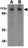
WB analysis of HepG2 cell lysate using GTX30881 LAMP2 antibody. Working concentration : (A) 1 and (B) 2 μg/ml


WB analysis of HepG2 cell lysate using GTX30881 LAMP2 antibody. Working concentration : (A) 1 and (B) 2 μg/ml
LAMP2 antibody
GTX30881
ApplicationsImmunoFluorescence, Western Blot, ELISA, ImmunoCytoChemistry, ImmunoHistoChemistry, ImmunoHistoChemistry Paraffin
Product group Antibodies
ReactivityHuman, Mouse
TargetLAMP2
Overview
- SupplierGeneTex
- Product NameLAMP2 antibody
- Delivery Days Customer9
- Application Supplier NoteWB: 1 - 2 microg/mL. ICC/IF: 10 microg/mL. *Optimal dilutions/concentrations should be determined by the researcher.Not tested in other applications.
- ApplicationsImmunoFluorescence, Western Blot, ELISA, ImmunoCytoChemistry, ImmunoHistoChemistry, ImmunoHistoChemistry Paraffin
- CertificationResearch Use Only
- ClonalityPolyclonal
- Concentration1 mg/ml
- ConjugateUnconjugated
- Gene ID3920
- Target nameLAMP2
- Target descriptionlysosomal associated membrane protein 2
- Target synonymsCD107b, DND, LAMP-2, LAMPB, LGP-96, LGP110, lysosome-associated membrane glycoprotein 2, CD107 antigen-like family member B
- HostRabbit
- IsotypeIgG
- Protein IDP13473
- Protein NameLysosome-associated membrane glycoprotein 2
- Scientific DescriptionAutophagy, the process of bulk degradation of cellular proteins through an autophagosomic-lysosomal pathway is important for normal growth control and may be defective in tumor cells. It is involved in the preservation of cellular nutrients under starvation conditions as well as the normal turnover of cytosolic components (1,2) and is negatively regulated by TOR (Target of rapamycin) (3). LAMP-2, a highly glycosylated protein associated with the lysosome (4), has recently been shown to be important in autophagy as mice deficient in this protein failed to convert autophagic vacuoles into vacuoles (5) leading to impaired degradation of long-lived proteins. This correlates with the finding that human LAMP-2 deficiency causing DanonOs disease is associated with the accumulation of autophagic material in striated myocytes (6). LAMP-2 exists in multiple isoforms (7).
- ReactivityHuman, Mouse
- Storage Instruction-20°C or -80°C,2°C to 8°C
- UNSPSC41116161
References
- SNX10 (sorting nexin 10) inhibits colorectal cancer initiation and progression by controlling autophagic degradation of SRC. Zhang S et al., 2019 Jul 4, AutophagyRead this paper
Datasheet
Related products
Product group Antibodies
Anti-LAMP2 AntibodyA99630
ApplicationsWestern Blot, ELISA, ImmunoHistoChemistry
ReactivityHuman
- SizePrice
Product group Antibodies
Anti-LAMP2 Antibody144-65436
ApplicationsWestern Blot, ImmunoHistoChemistry
ReactivityHuman
TargetLAMP2
- SizePrice

Product group Antibodies
Anti-LAMP2 Antibody Picoband(r)A01573-3-CARRIER-FREE
ApplicationsWestern Blot, ELISA
ReactivityHuman
TargetLAMP2
- SizePrice
Product group Antibodies
References
LAMP2 Polyclonal AntibodyBS-2379R
ApplicationsFlow Cytometry, ImmunoFluorescence, Western Blot, ELISA, ImmunoCytoChemistry, ImmunoHistoChemistry, ImmunoHistoChemistry Frozen, ImmunoHistoChemistry Paraffin
ReactivityHuman, Mouse, Rat
TargetLAMP2
- SizePrice
Product group Antibodies
LAMP2 AntibodyCSB-PA012740EA01HU
ApplicationsImmunoFluorescence, Western Blot, ELISA, ImmunoHistoChemistry
ReactivityHuman, Mouse
TargetLAMP2
- SizePrice
Product group Antibodies
Lamp2 Polyclonal AntibodyCAC07012
ApplicationsImmunoFluorescence, Western Blot, ELISA, ImmunoHistoChemistry
ReactivityMouse
TargetLAMP2
- SizePrice
Product group Antibodies

LAMP2 / CD107b AntibodyLS-C400703
ApplicationsWestern Blot, ELISA, ImmunoHistoChemistry
ReactivityHuman
TargetLAMP2
- SizePrice
Product group Antibodies
LAMP2A antibodyGTX132655
ApplicationsWestern Blot
ReactivityHuman
TargetLAMP2
- SizePrice
Product group Antibodies
References
LAMP2 antibody [GL2A7]GTX13524
ApplicationsFlow Cytometry, ImmunoFluorescence, ImmunoPrecipitation, Western Blot, ImmunoCytoChemistry, ImmunoHistoChemistry, ImmunoHistoChemistry Paraffin
ReactivityHuman, Mouse, Rabbit
TargetLAMP2
- SizePrice