

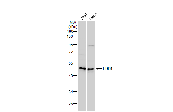
Various whole cell extracts (30 μg) were separated by 10% SDS-PAGE, and the membrane was blotted with LDB1 antibody [N2C3] (GTX104531) diluted at 1:1000. The HRP-conjugated anti-rabbit IgG antibody (GTX213110-01) was used to detect the primary antibody.
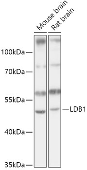
 A: Mouse brain 10% SDS PAGE GTX104531 diluted at 1:1000 The HRP-conjugated anti-rabbit IgG antibody (GTX213110-01) was used to detect the primary antibody.")
 antibody at 1:100 dilution.
Antigen Retrieval: Trilogy? (EDTA based, pH 8.0) buffer, 15min")
![LDB1 antibody [N2C3] detects LDB1 protein at nucleus by immunofluorescent analysis. Sample: HeLa cells were fixed in 4% paraformaldehyde at RT for 15 min. Green: LDB1 protein stained by LDB1 antibody [N2C3] (GTX104531) diluted at 1:500. Blue: Hoechst 33342 staining. Scale bar = 10 μm.](https://www.genetex.com/upload/website/prouct_img/normal/GTX104531/GTX104531_40709_20150513_IFA_w_23060120_173.webp "LDB1 antibody [N2C3] detects LDB1 protein at nucleus by immunofluorescent analysis. Sample: HeLa cells were fixed in 4% paraformaldehyde at RT for 15 min. Green: LDB1 protein stained by LDB1 antibody [N2C3] (GTX104531) diluted at 1:500. Blue: Hoechst 33342 staining. Scale bar = 10 μm.")
![LDB1 antibody [N2C3] detects LDB1 protein at nucleus on mouse muscle by immunohistochemical analysis. Sample: Paraffin-embedded mouse muscle. LDB1 antibody [N2C3] (GTX104531) dilution: 1:500.
Antigen Retrieval: Trilogy? (EDTA based, pH 8.0) buffer, 15min](https://www.genetex.com/upload/website/prouct_img/normal/GTX104531/GTX104531_41073_IHC_M_w_23060120_717.webp "LDB1 antibody [N2C3] detects LDB1 protein at nucleus on mouse muscle by immunohistochemical analysis. Sample: Paraffin-embedded mouse muscle. LDB1 antibody [N2C3] (GTX104531) dilution: 1:500.
Antigen Retrieval: Trilogy? (EDTA based, pH 8.0) buffer, 15min")
![Wild-type (WT) and LDB1 knockout (KO) HeLa cell extracts (30 μg) were separated by 10% SDS-PAGE, and the membrane was blotted with LDB1 antibody [N2C3] (GTX104531) diluted at 1:1000. The HRP-conjugated anti-rabbit IgG antibody (GTX213110-01) was used to detect the primary antibody, and the signal was developed with Trident ECL plus-Enhanced.](https://www.genetex.com/upload/website/prouct_img/normal/GTX104531/GTX104531_41766_20181109_WB_KO_watermark_w_23060120_703.webp "Wild-type (WT) and LDB1 knockout (KO) HeLa cell extracts (30 μg) were separated by 10% SDS-PAGE, and the membrane was blotted with LDB1 antibody [N2C3] (GTX104531) diluted at 1:1000. The HRP-conjugated anti-rabbit IgG antibody (GTX213110-01) was used to detect the primary antibody, and the signal was developed with Trident ECL plus-Enhanced.")
 12% SDS-PAGE The immunoprecipitated LDB1 protein was detected by LDB1antibody (GTX104531) diluted at 1:1000. EasyBlot anti-rabbit IgG (GTX221666-01) was used as a secondary reagent.")
![LDB1 antibody [N2C3] detects LDB1 protein at nucleus on rat middle brain by immunohistochemical analysis. Sample: Paraffin-embedded rat middle brain. LDB1 antibody [N2C3] (GTX104531) dilution: 1:500.
Antigen Retrieval: Trilogy? (EDTA based, pH 8.0) buffer, 15min](https://www.genetex.com/upload/website/prouct_img/normal/GTX104531/GTX104531_41073_IHC_R_w_23060120_549.webp "LDB1 antibody [N2C3] detects LDB1 protein at nucleus on rat middle brain by immunohistochemical analysis. Sample: Paraffin-embedded rat middle brain. LDB1 antibody [N2C3] (GTX104531) dilution: 1:500.
Antigen Retrieval: Trilogy? (EDTA based, pH 8.0) buffer, 15min")
![Various whole cell extracts (30 μg) were separated by 10% SDS-PAGE, and the membrane was blotted with LDB1 antibody [N2C3] (GTX104531) diluted at 1:1000. The HRP-conjugated anti-rabbit IgG antibody (GTX213110-01) was used to detect the primary antibody. Corresponding RNA expression data for the same cell lines are based on Human Protein Atlas program.](https://www.genetex.com/upload/website/prouct_img/normal/GTX104531/GTX104531_45582_20241108_WB_TPM_watermark_25070323_703.webp "Various whole cell extracts (30 μg) were separated by 10% SDS-PAGE, and the membrane was blotted with LDB1 antibody [N2C3] (GTX104531) diluted at 1:1000. The HRP-conjugated anti-rabbit IgG antibody (GTX213110-01) was used to detect the primary antibody. Corresponding RNA expression data for the same cell lines are based on Human Protein Atlas program.")
![IMR32 whole cell and nuclear extracts (30 μg) were separated by 10% SDS-PAGE, and the membrane was blotted with LDB1 antibody [N2C3] (GTX104531) diluted at 1:1000. The HRP-conjugated anti-rabbit IgG antibody (GTX213110-01) was used to detect the primary antibody.](https://www.genetex.com/upload/website/prouct_img/normal/GTX104531/GTX104531_45582_20250117_WB_Fraction_25070323_737.webp "IMR32 whole cell and nuclear extracts (30 μg) were separated by 10% SDS-PAGE, and the membrane was blotted with LDB1 antibody [N2C3] (GTX104531) diluted at 1:1000. The HRP-conjugated anti-rabbit IgG antibody (GTX213110-01) was used to detect the primary antibody.")
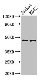
Various whole cell extracts (30 μg) were separated by 10% SDS-PAGE, and the membrane was blotted with LDB1 antibody [N2C3] (GTX104531) diluted at 1:1000. The HRP-conjugated anti-rabbit IgG antibody (GTX213110-01) was used to detect the primary antibody.
LDB1 antibody [N2C3]
GTX104531
ApplicationsImmunoFluorescence, ImmunoPrecipitation, Western Blot, ImmunoCytoChemistry, ImmunoHistoChemistry, ImmunoHistoChemistry Paraffin
Product group Antibodies
ReactivityHuman, Mouse, Rat
TargetLDB1
Overview
- SupplierGeneTex
- Product NameLDB1 antibody [N2C3]
- Delivery Days Customer9
- Application Supplier NoteWB: 1:500-1:3000. ICC/IF: 1:100-1:1000. IHC-P: 1:100-1:1000. IP: 1:100-1:500. *Optimal dilutions/concentrations should be determined by the researcher.Not tested in other applications.
- ApplicationsImmunoFluorescence, ImmunoPrecipitation, Western Blot, ImmunoCytoChemistry, ImmunoHistoChemistry, ImmunoHistoChemistry Paraffin
- CertificationResearch Use Only
- ClonalityPolyclonal
- Concentration0.59 mg/ml
- ConjugateUnconjugated
- Gene ID8861
- Target nameLDB1
- Target descriptionLIM domain binding 1
- Target synonymsCLIM-2, CLIM2, LDB-1, NLI, LIM domain-binding protein 1, LIM domain-binding factor CLIM2, LIM domain-binding factor-1, carboxy terminal LIM domain protein 2, carboxyl-terminal LIM domain-binding protein 2, nuclear LIM interactor
- HostRabbit
- IsotypeIgG
- Protein IDQ86U70
- Protein NameLIM domain-binding protein 1
- Scientific DescriptionBinds to the LIM domain of a wide variety of LIM domain-containing transcription factors. May regulate the transcriptional activity of LIM-containing proteins by determining specific partner interactions. May play a role in the development of motor neurons. Acts synergistically with LHX1/LIM1 in axis formation and activation of gene expression. Acts with LMO2 in the regulation of red blood cell development, maintaining erythroid precursors in an immature state.
- ReactivityHuman, Mouse, Rat
- Storage Instruction-20°C or -80°C,2°C to 8°C
- UNSPSC41116161
Datasheet
Related products
Product group Antibodies
Anti-LDB1 AntibodyA307494
ApplicationsWestern Blot, ImmunoHistoChemistry
ReactivityHuman, Mouse, Rat
- SizePrice
Product group Antibodies
Anti-LDB1 Antibody144-65009
ApplicationsWestern Blot, ImmunoHistoChemistry
ReactivityHuman, Mouse, Rat
TargetLDB1
- SizePrice
Product group Antibodies

LDB1 / CLIM2 AntibodyLS-C830768
ApplicationsELISA, ImmunoHistoChemistry
ReactivityHuman, Mouse
TargetLDB1
- SizePrice
Product group Antibodies
LDB1 AntibodyCSB-PA771433LA01HU
ApplicationsImmunoFluorescence, Western Blot, ELISA, ImmunoHistoChemistry
ReactivityHuman
TargetLDB1
- SizePrice
Product group Antibodies
LDB1 Polyclonal AntibodyCAC14999
ApplicationsImmunoFluorescence, Western Blot, ELISA, ImmunoHistoChemistry
TargetLDB1
- SizePrice

Product group Antibodies
Anti-LDB1 AntibodyHPA034488
ApplicationsWestern Blot, ChIP Chromatin ImmunoPrecipitation, ImmunoCytoChemistry, ImmunoHistoChemistry
ReactivityHuman
TargetLDB1
- SizePrice