

LMO2 (B-Cell Marker)(rLMO2/1971), CF594 conjugate, 0.1mg/mL [26628-22-8]
BNC943806
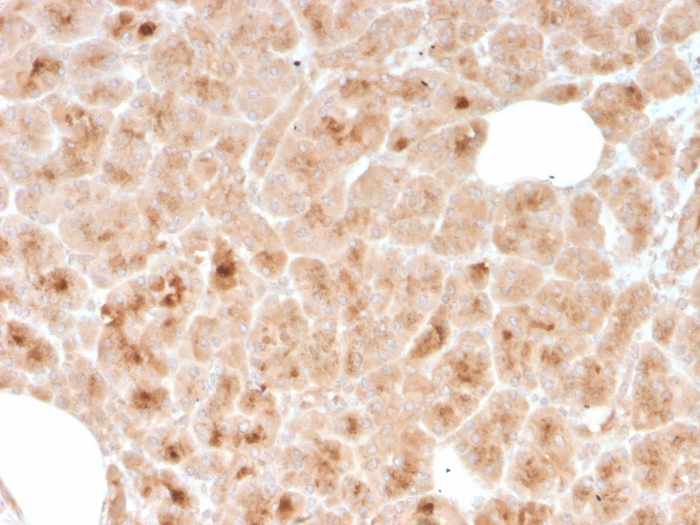
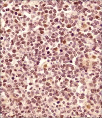
ApplicationsImmunoHistoChemistry, ImmunoHistoChemistry Paraffin
Product group Antibodies
ReactivityHuman
TargetLMO2
Overview
- SupplierBiotium
- Product NameLMO2 (B-Cell Marker)(rLMO2/1971), CF594 conjugate, 0.1mg/mL [26628-22-8]
- Delivery Days Customer9
- ApplicationsImmunoHistoChemistry, ImmunoHistoChemistry Paraffin
- CAS Number26628-22-8
- CertificationResearch Use Only
- ClonalityMonoclonal
- Clone IDrLMO2/1971
- Concentration0.1 mg/ml
- ConjugateOther Conjugate
- Gene ID4005
- Target nameLMO2
- Target descriptionLIM domain only 2
- Target synonymsLMO-2, RBTN2, RBTNL1, RHOM2, TTG2, rhombotin-2, LIM domain only protein 2, T-cell translocation gene 2, T-cell translocation protein 2, cysteine-rich protein TTG-2, rhombotin-like 1
- HostMouse
- IsotypeIgG1
- Protein IDP25791
- Protein NameRhombotin-2
- Scientific DescriptionThe LMO2 protein has a central and crucial role in hematopoietic development and is highly conserved. It has a particular function in normal and lymphatic endothelial cells involving the regulation of angiogenesis and lymph-angiogenesis. Immunohistochemical studies have also demonstrated expression of LMO2 in both normal germinal center B-cells and germinal center-derived B-cell lymphomas, including follicular lymphoma and diffuse large B-cell lymphoma. The use of anti-LMO2 is valuable as a tool in the identification of lymphomas of B-cell origin. LMO2 is useful in differentiating follicular lymphoma (LMO2 ) from nodal marginal zone lymphoma (LM02-). It also is positive in Hodgkins and Burkitts lymphomas. Primary antibodies are available purified, or with a selection of fluorescent CF® Dyes and other labels. CF® Dyes offer exceptional brightness and photostability. Note: Conjugates of blue fluorescent dyes like CF®405S and CF®405M are not recommended for detecting low abundance targets, because blue dyes have lower fluorescence and can give higher non-specific background than other dye colors.
- SourceAnimal
- ReactivityHuman
- Storage Instruction2°C to 8°C,RT
- UNSPSC41116161
MSDS
Related products
Product group Antibodies
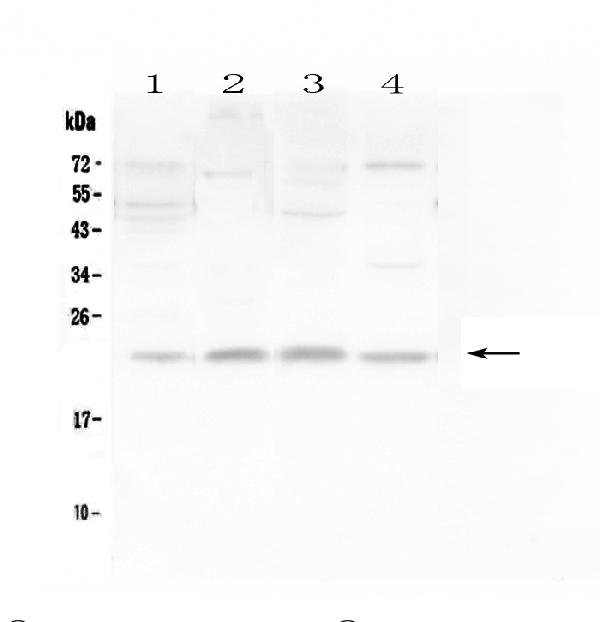
Anti-LMO2 Antibody Picoband(r)A03502-1-CARRIER-FREE
ApplicationsWestern Blot
ReactivityHuman, Mouse, Rat
TargetLMO2
- SizePrice
Product group Antibodies
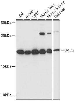
Anti-LMO2 Antibody144-01903
ApplicationsWestern Blot, ImmunoHistoChemistry
ReactivityHuman, Mouse, Rat
TargetLMO2
- SizePrice
Product group Antibodies
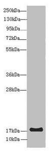
Anti-LMO2 AntibodyA13732
ApplicationsWestern Blot
ReactivityHuman, Mouse, Rat
- SizePrice
Product group Antibodies

LMO2 AntibodyLS-C747769
ApplicationsWestern Blot
ReactivityHuman, Mouse
TargetLMO2
- SizePrice
Product group Antibodies
LMO2 Recombinant AntibodyBSM-60228R
ApplicationsFlow Cytometry, Western Blot
ReactivityHuman
TargetLMO2
- SizePrice
Product group Antibodies
LMO2 AntibodyCSB-PA013009HA01HU
ApplicationsWestern Blot, ELISA, ImmunoHistoChemistry
ReactivityHuman, Mouse
TargetLMO2
- SizePrice
Product group Antibodies
Lmo2 Polyclonal AntibodyCAC07614
ApplicationsWestern Blot, ELISA, ImmunoHistoChemistry
ReactivityMouse
TargetLMO2
- SizePrice

Product group Antibodies
Anti-LMO2 AntibodyHPA029285
ApplicationsImmunoCytoChemistry
ReactivityHuman
TargetLMO2
- SizePrice
Product group Antibodies
LMO2 antibodyGTX48597
ApplicationsWestern Blot, ChIP Chromatin ImmunoPrecipitation, ImmunoHistoChemistry, ImmunoHistoChemistry Paraffin
ReactivityBovine, Human, Mouse, Rat, Zebra Fish
TargetLMO2
- SizePrice