

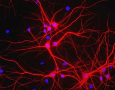
ICC/IF analysis of mixed neuron/glia cell cultures using GTX30663 MAP2 antibody. The perikarya and dendrites of neurons are strongly and specifically stained with the MAP2 antibody, while the axons of the neurons and the processes of all other cell types in these cultures (astrocytes, oligodendrocytes, microglia, endothelia and fibroblasts) are all negative.

ICC/IF analysis of mixed neuron/glia cell cultures using GTX30663 MAP2 antibody. The perikarya and dendrites of neurons are strongly and specifically stained with the MAP2 antibody, while the axons of the neurons and the processes of all other cell types in these cultures (astrocytes, oligodendrocytes, microglia, endothelia and fibroblasts) are all negative.
MAP2 antibody
GTX30663
ApplicationsImmunoFluorescence, Western Blot, ImmunoCytoChemistry, ImmunoHistoChemistry, ImmunoHistoChemistry Frozen
Product group Antibodies
ReactivityBovine, Human, Mouse, Rat
Overview
- SupplierGeneTex
- Product NameMAP2 antibody
- Delivery Days Customer9
- Application Supplier NoteWB: 1:10000 - 1:20000. ICC/IF: 1:5000 - 1:10000. IHC-Fr: 1:1000 - 1:5000. IHC: 1:1000 - 1:5000. *Optimal dilutions/concentrations should be determined by the researcher.Not tested in other applications.
- ApplicationsImmunoFluorescence, Western Blot, ImmunoCytoChemistry, ImmunoHistoChemistry, ImmunoHistoChemistry Frozen
- CertificationResearch Use Only
- ClonalityPolyclonal
- ConjugateUnconjugated
- HostChicken
- IsotypeIgY
- Scientific DescriptionMicrotubules are 25nm diameter protein rods found in most kinds of eukarytic cells. They are polymerized from a dimeric subunit made of one a subunit and one b tubulin subunit. Microtubules are associated with a family of proteins called microtubule associated proteins (MAPs), which includes the protein t (tau) and a group of proteins referred to as MAP1, MAP2, MAP3, MAP4 and MAP5. MAP2 is made up of two ~280kDa apparent molecular weight bands referred to as MAP2a and MAP2b. A third lower molecular weight form, usually called MAP2c, corresponds to a pair of protein bands running at ~70kDa on SDS-PAGE gels. Antibodies to MAP2 are excellent markers on neuronal cells, their perikarya and neuronal dendrites. In contrast t (tau) is found predominantly in neuronal axons.
- ReactivityBovine, Human, Mouse, Rat
- Storage Instruction-20°C or -80°C,2°C to 8°C
- UNSPSC41116161
References
- Netrin-1 promotes excitatory synaptogenesis between cortical neurons by initiating synapse assembly. Goldman JS et al., 2013 Oct 30, J NeurosciRead this paper
- Molecular characterization of the mouse superior lateral parabrachial nucleus through expression of the transcription factor Runx1. Zagami CJ et al., 2010 Nov 11, PLoS OneRead this paper
Datasheet
Related products
Product group Antibodies
Map2 AntibodyCSB-PA130852
ApplicationsELISA, ImmunoHistoChemistry
ReactivityHuman, Mouse, Rat
TargetMap2
- SizePrice
![MAP2 antibody [HL1655] detects MAP2 protein in dendrite/axon, by immunofluorescent analysis. Sample: DIV9 rat E18 primary hippocampal neuron cells were fixed in 4% paraformaldehyde at RT for 15 min. Green: MAP2 stained by MAP2 antibody [HL1655] (GTX637253) diluted at 1:250. Red: Tau, an axon marker, stained by Tau antibody [GT287] (GTX634809) diluted at 1:500. Blue: Fluoroshield with DAPI (GTX30920).](https://www.genetex.com/upload/website/prouct_img/normal/GTX637253/GTX637253_T-44753_20221209_ICC_IF_R_22122018_593.webp)
Product group Antibodies
MAP2 antibody [HL1655]GTX637253
ApplicationsImmunoFluorescence, ImmunoCytoChemistry, ImmunoHistoChemistry, ImmunoHistoChemistry Paraffin
ReactivityMouse, Rat
TargetMap2
- SizePrice
![MAP2 antibody [HL1656] detects MAP2 protein in dendrites, but not in axons, by immunofluorescent analysis. Sample: DIV9 rat E18 primary hippocampal neuron cells were fixed in 4% paraformaldehyde at RT for 15 min. Green: MAP2 stained by MAP2 antibody [HL1656] (GTX637254) diluted at 1:250. Red: Tau, an axon marker, stained by Tau antibody [GT287] (GTX634809) diluted at 1:500. Blue: Fluoroshield with DAPI (GTX30920).](https://www.genetex.com/upload/website/prouct_img/normal/GTX637254/GTX637254_T-44753_20221209_ICC_IF_R_22122018_267.webp)
Product group Antibodies
MAP2 antibody [HL1656]GTX637254
ApplicationsImmunoFluorescence, ImmunoCytoChemistry, ImmunoHistoChemistry, ImmunoHistoChemistry Paraffin
ReactivityMouse, Rat
TargetMap2
- SizePrice
![MAP2 antibody [GT378] detects MAP2 protein in dendrites, but not in axons, by immunofluorescent analysis. Sample: DIV10 rat E18 primary cortical neuron cells were fixed in 4% paraformaldehyde at RT for 15 min. Green: Dendrite marker MAP2 stained by MAP2 antibody [GT378] (GTX634471) diluted at 1:1000. Red: Axon marker Tau, stained by Tau antibody (GTX130462) diluted at 1:500. Blue: Fluoroshield with DAPI (GTX30920).](https://www.genetex.com/upload/website/prouct_img/normal/GTX634471/GTX634471_43264_20180516_ICC_IF_R_w_23061202_424.webp)
Product group Antibodies
MAP2 antibody [GT378]GTX634471
ApplicationsImmunoFluorescence, Western Blot, ImmunoCytoChemistry, ImmunoHistoChemistry, ImmunoHistoChemistry Paraffin
ReactivityMouse, Rat
TargetMap2
- SizePrice
![MAP2 antibody [GT925] detects MAP2 protein at cytoplasm by immunohistochemical analysis. Sample: Paraffin-embedded mouse cerebellum. MAP2 stained by MAP2 antibody [GT925] (GTX634473) diluted at 1:2000.
Antigen Retrieval: Citrate buffer, pH 6.0, 15 min](https://www.genetex.com/upload/website/prouct_img/normal/GTX634473/GTX634473_43178_20180803_IHC-P_M_w_23061202_486.webp)
Product group Antibodies
MAP2 antibody [GT925]GTX634473
ApplicationsImmunoFluorescence, Western Blot, ImmunoCytoChemistry, ImmunoHistoChemistry, ImmunoHistoChemistry Paraffin
ReactivityMouse, Rat
TargetMap2
- SizePrice
Product group Antibodies
MAP2 antibodyGTX32712
ApplicationsWestern Blot
ReactivityHuman, Mouse
TargetMap2
- SizePrice
Product group Antibodies
MAP2 antibodyGTX133109
ApplicationsImmunoFluorescence, Western Blot, ImmunoCytoChemistry, ImmunoHistoChemistry, ImmunoHistoChemistry Frozen, ImmunoHistoChemistry Paraffin
ReactivityFish, Human, Mouse, Rat
TargetMap2
- SizePrice
![MAP2 antibody detects MAP2 protein in dendrites, but not in axons, by immunofluorescent analysis. Sample: DIV9 rat E18 primary cortical neurons were fixed in 4% paraformaldehyde at RT for 15 min. Grenn: MAP2 protein stained by MAP2 antibody (GTX133110) diluted at 1:500. Red (Top right): Dendrites, stained by MAP2 antibody [HM-2] (GTX11267) diluted at 1:1000. Red (Bottom right): Axons, stained by Tau antibody (GTX49353) diluted at 1:1000. Blue: Fluoroshield with DAPI (GTX30920).](https://www.genetex.com/upload/website/prouct_img/normal/GTX133110/GTX133110_42592_20170705_IFA_R_3_w_23060523_118.webp)
Product group Antibodies
MAP2 antibodyGTX133110
ApplicationsImmunoFluorescence, Western Blot, ImmunoCytoChemistry, ImmunoHistoChemistry, ImmunoHistoChemistry Frozen
ReactivityMouse, Rat
TargetMap2
- SizePrice
Product group Antibodies
ApplicationsImmunoPrecipitation, Western Blot, ImmunoCytoChemistry, ImmunoHistoChemistry
ReactivityMouse, Rat
TargetMap2
- SizePrice