

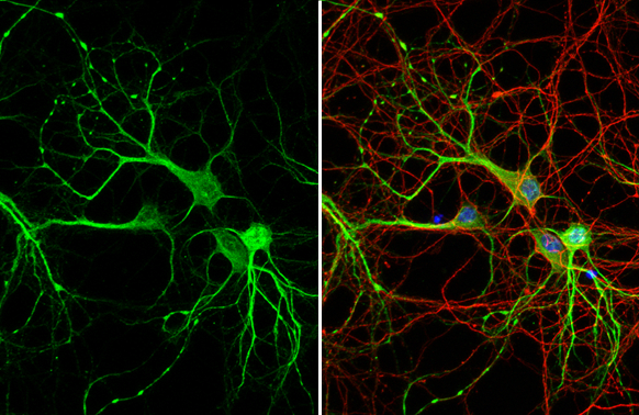
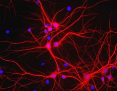
MAP2 antibody [HL1655] detects MAP2 protein in dendrite/axon, by immunofluorescent analysis. Sample: DIV9 rat E18 primary hippocampal neuron cells were fixed in 4% paraformaldehyde at RT for 15 min. Green: MAP2 stained by MAP2 antibody [HL1655] (GTX637253) diluted at 1:250. Red: Tau, an axon marker, stained by Tau antibody [GT287] (GTX634809) diluted at 1:500. Blue: Fluoroshield with DAPI (GTX30920).
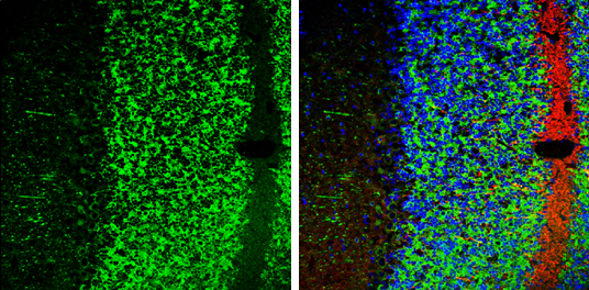
![MAP2 antibody [HL1655] detects MAP2 protein by immunohistochemical analysis. Sample: Paraffin-embedded mouse cerebellum. Green: MAP2 stained by MAP2 antibody [HL1655] (GTX637253) diluted at 1:100. Red: beta Tubulin 3/ Tuj1, a Cytoskeleton marker, stained by beta Tubulin 3/ Tuj1 antibody [GT11710] (GTX631836) diluted at 1:500. Blue: Fluoroshield with DAPI (GTX30920). Antigen Retrieval: Citrate buffer, pH 6.0, 15 min](https://www.genetex.com/upload/website/prouct_img/normal/GTX637253/GTX637253_44935_20230303_IHC-P_M_1_23031402_205.webp "MAP2 antibody [HL1655] detects MAP2 protein by immunohistochemical analysis. Sample: Paraffin-embedded mouse cerebellum. Green: MAP2 stained by MAP2 antibody [HL1655] (GTX637253) diluted at 1:100. Red: beta Tubulin 3/ Tuj1, a Cytoskeleton marker, stained by beta Tubulin 3/ Tuj1 antibody [GT11710] (GTX631836) diluted at 1:500. Blue: Fluoroshield with DAPI (GTX30920). Antigen Retrieval: Citrate buffer, pH 6.0, 15 min")
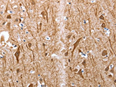
![MAP2 antibody [HL1655] detects MAP2 protein by immunohistochemical analysis. Sample: Paraffin-embedded rat tissues. MAP2 stained by MAP2 antibody [HL1655] (GTX637253) diluted at 1:100. Antigen Retrieval: Citrate buffer, pH 6.0, 15 min](https://www.genetex.com/upload/website/prouct_img/normal/GTX637253/GTX637253_44935_20230303_IHC-P_multiple_R_23031402_526.webp "MAP2 antibody [HL1655] detects MAP2 protein by immunohistochemical analysis. Sample: Paraffin-embedded rat tissues. MAP2 stained by MAP2 antibody [HL1655] (GTX637253) diluted at 1:100. Antigen Retrieval: Citrate buffer, pH 6.0, 15 min")
![MAP2 antibody [HL1655] detects MAP2 protein by immunohistochemical analysis. Sample: Paraffin-embedded mouse tissues. MAP2 stained by MAP2 antibody [HL1655] (GTX637253) diluted at 1:100. Antigen Retrieval: Citrate buffer, pH 6.0, 15 min](https://www.genetex.com/upload/website/prouct_img/normal/GTX637253/GTX637253_45670_20250314_IHC-P_M_Multiple_RPKM_25032719_787.webp "MAP2 antibody [HL1655] detects MAP2 protein by immunohistochemical analysis. Sample: Paraffin-embedded mouse tissues. MAP2 stained by MAP2 antibody [HL1655] (GTX637253) diluted at 1:100. Antigen Retrieval: Citrate buffer, pH 6.0, 15 min")
MAP2 antibody [HL1655] detects MAP2 protein in dendrite/axon, by immunofluorescent analysis. Sample: DIV9 rat E18 primary hippocampal neuron cells were fixed in 4% paraformaldehyde at RT for 15 min. Green: MAP2 stained by MAP2 antibody [HL1655] (GTX637253) diluted at 1:250. Red: Tau, an axon marker, stained by Tau antibody [GT287] (GTX634809) diluted at 1:500. Blue: Fluoroshield with DAPI (GTX30920).
MAP2 antibody [HL1655]
GTX637253
ApplicationsImmunoFluorescence, ImmunoCytoChemistry, ImmunoHistoChemistry, ImmunoHistoChemistry Paraffin
Product group Antibodies
ReactivityMouse, Rat
TargetMap2
Overview
- SupplierGeneTex
- Product NameMAP2 antibody [HL1655]
- Delivery Days Customer9
- Application Supplier NoteIHC-P: 1:100-1:1000. *Optimal dilutions/concentrations should be determined by the researcher.Not tested in other applications.
- ApplicationsImmunoFluorescence, ImmunoCytoChemistry, ImmunoHistoChemistry, ImmunoHistoChemistry Paraffin
- CertificationResearch Use Only
- ClonalityMonoclonal
- Clone IDHL1655
- Concentration1 mg/ml
- ConjugateUnconjugated
- Gene ID17756
- Target nameMap2
- Target descriptionmicrotubule-associated protein 2
- Target synonymsG1-397-34, MAP-2, Mtap-2, Mtap2, repro4, microtubule-associated protein 2
- HostRabbit
- IsotypeIgG
- Protein IDP20357
- Protein NameMicrotubule-associated protein 2
- Scientific DescriptionEnables microtubule binding activity. Involved in microtubule cytoskeleton organization; negative regulation of axon extension; and regulation of protein localization. Acts upstream of or within several processes, including establishment of cell polarity; microtubule bundle formation; and neuron projection development. Located in several cellular components, including axon; dendrite; and proximal neuron projection. Is expressed in several structures, including back skin; central nervous system; peripheral nervous system ganglion; retina; and trigeminal nerve. Orthologous to human MAP2 (microtubule associated protein 2). [provided by Alliance of Genome Resources, Apr 2022]
- ReactivityMouse, Rat
- Storage Instruction-20°C or -80°C,2°C to 8°C
- UNSPSC41116161
Datasheet
Related products
Product group Antibodies
Map2 AntibodyCSB-PA130852
ApplicationsELISA, ImmunoHistoChemistry
ReactivityHuman, Mouse, Rat
TargetMap2
- SizePrice
![MAP2 antibody [HL1656] detects MAP2 protein in dendrites, but not in axons, by immunofluorescent analysis. Sample: DIV9 rat E18 primary hippocampal neuron cells were fixed in 4% paraformaldehyde at RT for 15 min. Green: MAP2 stained by MAP2 antibody [HL1656] (GTX637254) diluted at 1:250. Red: Tau, an axon marker, stained by Tau antibody [GT287] (GTX634809) diluted at 1:500. Blue: Fluoroshield with DAPI (GTX30920).](https://www.genetex.com/upload/website/prouct_img/normal/GTX637254/GTX637254_T-44753_20221209_ICC_IF_R_22122018_267.webp)
Product group Antibodies
MAP2 antibody [HL1656]GTX637254
ApplicationsImmunoFluorescence, ImmunoCytoChemistry, ImmunoHistoChemistry, ImmunoHistoChemistry Paraffin
ReactivityMouse, Rat
TargetMap2
- SizePrice
![MAP2 antibody [GT378] detects MAP2 protein in dendrites, but not in axons, by immunofluorescent analysis. Sample: DIV10 rat E18 primary cortical neuron cells were fixed in 4% paraformaldehyde at RT for 15 min. Green: Dendrite marker MAP2 stained by MAP2 antibody [GT378] (GTX634471) diluted at 1:1000. Red: Axon marker Tau, stained by Tau antibody (GTX130462) diluted at 1:500. Blue: Fluoroshield with DAPI (GTX30920).](https://www.genetex.com/upload/website/prouct_img/normal/GTX634471/GTX634471_43264_20180516_ICC_IF_R_w_23061202_424.webp)
Product group Antibodies
MAP2 antibody [GT378]GTX634471
ApplicationsImmunoFluorescence, Western Blot, ImmunoCytoChemistry, ImmunoHistoChemistry, ImmunoHistoChemistry Paraffin
ReactivityMouse, Rat
TargetMap2
- SizePrice
![MAP2 antibody [GT925] detects MAP2 protein at cytoplasm by immunohistochemical analysis. Sample: Paraffin-embedded mouse cerebellum. MAP2 stained by MAP2 antibody [GT925] (GTX634473) diluted at 1:2000.
Antigen Retrieval: Citrate buffer, pH 6.0, 15 min](https://www.genetex.com/upload/website/prouct_img/normal/GTX634473/GTX634473_43178_20180803_IHC-P_M_w_23061202_486.webp)
Product group Antibodies
MAP2 antibody [GT925]GTX634473
ApplicationsImmunoFluorescence, Western Blot, ImmunoCytoChemistry, ImmunoHistoChemistry, ImmunoHistoChemistry Paraffin
ReactivityMouse, Rat
TargetMap2
- SizePrice
Product group Antibodies
MAP2 antibodyGTX32712
ApplicationsWestern Blot
ReactivityHuman, Mouse
TargetMap2
- SizePrice
Product group Antibodies
MAP2 antibodyGTX133109
ApplicationsImmunoFluorescence, Western Blot, ImmunoCytoChemistry, ImmunoHistoChemistry, ImmunoHistoChemistry Frozen, ImmunoHistoChemistry Paraffin
ReactivityFish, Human, Mouse, Rat
TargetMap2
- SizePrice
![MAP2 antibody detects MAP2 protein in dendrites, but not in axons, by immunofluorescent analysis. Sample: DIV9 rat E18 primary cortical neurons were fixed in 4% paraformaldehyde at RT for 15 min. Grenn: MAP2 protein stained by MAP2 antibody (GTX133110) diluted at 1:500. Red (Top right): Dendrites, stained by MAP2 antibody [HM-2] (GTX11267) diluted at 1:1000. Red (Bottom right): Axons, stained by Tau antibody (GTX49353) diluted at 1:1000. Blue: Fluoroshield with DAPI (GTX30920).](https://www.genetex.com/upload/website/prouct_img/normal/GTX133110/GTX133110_42592_20170705_IFA_R_3_w_23060523_118.webp)
Product group Antibodies
MAP2 antibodyGTX133110
ApplicationsImmunoFluorescence, Western Blot, ImmunoCytoChemistry, ImmunoHistoChemistry, ImmunoHistoChemistry Frozen
ReactivityMouse, Rat
TargetMap2
- SizePrice
Product group Antibodies
References
MAP2 antibodyGTX30663
ApplicationsImmunoFluorescence, Western Blot, ImmunoCytoChemistry, ImmunoHistoChemistry, ImmunoHistoChemistry Frozen
ReactivityBovine, Human, Mouse, Rat
- SizePrice
Product group Antibodies
ApplicationsImmunoPrecipitation, Western Blot, ImmunoCytoChemistry, ImmunoHistoChemistry
ReactivityMouse, Rat
TargetMap2
- SizePrice