

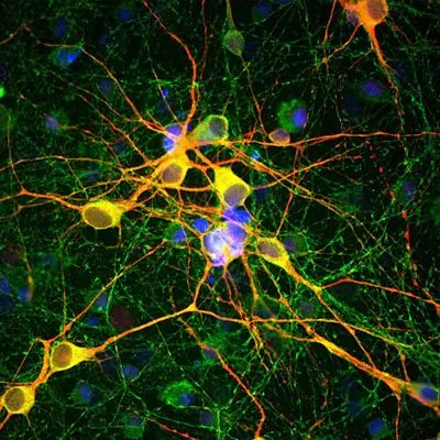
ICC/IF analysis of neuron-glial cell cultures from E20 rat using GTX82661 MAP2 antibody. The anti-MAP2 antibody stains dendrites and perikarya of neurons, while the anti-TAU antibody labels neuronal perikarya, dendrites and also axonal process. As a result perikarya and dendrites appears orange-yellow, since they contain both MAP2 and tau, while axons are green. Red : MAP2 Green : Tau Blue : DAPI Dilution : 1:10,000


ICC/IF analysis of neuron-glial cell cultures from E20 rat using GTX82661 MAP2 antibody. The anti-MAP2 antibody stains dendrites and perikarya of neurons, while the anti-TAU antibody labels neuronal perikarya, dendrites and also axonal process. As a result perikarya and dendrites appears orange-yellow, since they contain both MAP2 and tau, while axons are green. Red : MAP2 Green : Tau Blue : DAPI Dilution : 1:10,000
MAP2 antibody
GTX82661
ApplicationsImmunoFluorescence, Western Blot, ImmunoCytoChemistry, ImmunoHistoChemistry, Other Application
Product group Antibodies
ReactivityBovine, Human, Mouse, Rat
TargetMAP2
Overview
- SupplierGeneTex
- Product NameMAP2 antibody
- Delivery Days Customer9
- Application Supplier NoteWB: 1:20000. ICC/IF: 1:1000-1:20000. IHC: 1:2500-1:10000. *Optimal dilutions/concentrations should be determined by the researcher.Not tested in other applications.
- ApplicationsImmunoFluorescence, Western Blot, ImmunoCytoChemistry, ImmunoHistoChemistry, Other Application
- CertificationResearch Use Only
- ClonalityPolyclonal
- ConjugateUnconjugated
- Gene ID4133
- Target nameMAP2
- Target descriptionmicrotubule associated protein 2
- Target synonymsMAP-2, MAP2A, MAP2B, MAP2C, microtubule-associated protein 2
- HostChicken
- IsotypeIgY
- Protein IDP11137
- Protein NameMicrotubule-associated protein 2
- Scientific DescriptionThis gene encodes a protein that belongs to the microtubule-associated protein family. The proteins of this family are thought to be involved in microtubule assembly, which is an essential step in neurogenesis. The products of similar genes in rat and mouse are neuron-specific cytoskeletal proteins that are enriched in dentrites, implicating a role in determining and stabilizing dentritic shape during neuron development. A number of alternatively spliced variants encoding distinct isoforms have been described. [provided by RefSeq, Jan 2010]
- ReactivityBovine, Human, Mouse, Rat
- Storage Instruction-20°C or -80°C,2°C to 8°C
- UNSPSC41116161
References
- Stimulating VAPB-PTPIP51 ER-mitochondria tethering corrects FTD/ALS mutant TDP43 linked Ca(2+) and synaptic defects.Read this paper
- Functional neuronal circuitry and oscillatory dynamics in human brain organoids. Sharf T et al., 2022 Jul 29, Nat CommunRead this paper
- Bridging Integrator-1 protein loss in Alzheimers disease promotes synaptic tau accumulation and disrupts tau release. Glennon EB et al., 2020, Brain CommunRead this paper
Datasheet
Related products
Product group Antibodies

ApplicationsImmunoFluorescence, Western Blot, ImmunoCytoChemistry, ImmunoHistoChemistry
- SizePrice

Product group Antibodies
Anti-MAP2 Antibody Picoband(r)A01201-4-CARRIER-FREE
ApplicationsWestern Blot, ELISA, ImmunoHistoChemistry
ReactivityHuman, Mouse, Rat
TargetMAP2
- SizePrice
Product group Antibodies
Anti-Map2 Antibody144-61622
ApplicationsWestern Blot, ImmunoHistoChemistry
ReactivityHuman, Mouse, Rat
TargetMAP2
- SizePrice

Product group Antibodies
Anti-MAP2 AntibodyAMAB91375
ApplicationsWestern Blot, ImmunoCytoChemistry, ImmunoHistoChemistry
ReactivityHuman
TargetMAP2
- SizePrice
Product group Antibodies

MAP2 AntibodyLS-C813046
ApplicationsImmunoFluorescence, Western Blot
ReactivityBovine, Human, Mouse, Porcine, Rat
TargetMAP2
- SizePrice
Product group Antibodies
References
MAP2 Polyclonal AntibodyBS-1369R
ApplicationsFlow Cytometry, ImmunoFluorescence, Western Blot, ELISA, ImmunoCytoChemistry, ImmunoHistoChemistry, ImmunoHistoChemistry Frozen, ImmunoHistoChemistry Paraffin
ReactivityHuman, Mouse, Rat
TargetMAP2
- SizePrice
Product group Antibodies
MAP2 Monoclonal AntibodyCSB-MA000225
ApplicationsELISA, ImmunoHistoChemistry
ReactivityHuman, Mouse, Rat
TargetMAP2
- SizePrice
Product group Antibodies
ApplicationsWestern Blot, ELISA, ImmunoCytoChemistry, ImmunoHistoChemistry, ImmunoHistoChemistry Frozen, ImmunoHistoChemistry Paraffin
ReactivityMouse, Porcine, Rat
TargetMAP2
- SizePrice
![WB analysis of mouse brain tissue lysate and HeLa whole cell lysate using GTX03485 MAP2 antibody [1D7-B9-C8-F6]. Dilution : 1:1000](https://www.genetex.com/upload/website/prouct_img/normal/GTX03485/GTX03485_20220609_WB_164_w_23053123_780.webp)
Product group Antibodies
MAP2 antibody [1D7-B9-C8-F6]GTX03485
ApplicationsImmunoFluorescence, ImmunoPrecipitation, Western Blot, ImmunoCytoChemistry
ReactivityHuman, Mouse
TargetMAP2
- SizePrice
Product group Antibodies
References
MAP2 antibodyGTX85455
ApplicationsImmunoFluorescence, Western Blot, ImmunoCytoChemistry, ImmunoHistoChemistry
ReactivityHuman, Mouse, Rat
TargetMAP2
- SizePrice