

MCP2 antibody [35509.11]
GTX10391
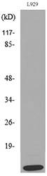
ApplicationsWestern Blot, ELISA, Neutralisation/Blocking
Product group Antibodies
ReactivityHuman
TargetCCL8
Overview
- SupplierGeneTex
- Product NameMCP2 antibody [35509.11]
- Delivery Days Customer9
- Application Supplier NoteWB: 1-2 microg/ml. ELISA: 2.0 microg/ml. Neutralizing/Inhibition: 1-4 microg/ml. *Optimal dilutions/concentrations should be determined by the researcher.Not tested in other applications.
- ApplicationsWestern Blot, ELISA, Neutralisation/Blocking
- CertificationResearch Use Only
- ClonalityMonoclonal
- Clone ID35509.11
- ConjugateUnconjugated
- Gene ID6355
- Target nameCCL8
- Target descriptionC-C motif chemokine ligand 8
- Target synonymsHC14, MCP-2, MCP2, SCYA10, SCYA8, C-C motif chemokine 8, chemokine (C-C motif) ligand 8, monocyte chemoattractant protein 2, monocyte chemotactic protein 2, small inducible cytokine subfamily A (Cys-Cys), member 8 (monocyte chemotactic protein 2), small-inducible cytokine A8
- HostMouse
- IsotypeIgG1
- Protein IDP80075
- Protein NameC-C motif chemokine 8
- Scientific DescriptionMCP2 and MCP3 are members of the C-C, or beta chemokine class. Other chemokines in this group include C10, Eotaxin, HCC1, I309, JE, MCP1, MIP1 alpha, MIP1 beta and RANTES. These chemokines act primarily as chemoattractants and antibody does not cross-react with other activate monocytes, dendritic cells, T lymphocytes, natural killer cells, B lymphocytes, basophils and eosinophils. MCP2 and MCP3 were originally identified as monocyte chemotactic proteins produced by human MG63 osteosacrcoma cells. MCP2 and MCP3 are approximately 9 kD polypeptides of 76 amino acids. MCP2 shares 62% amino acid sequence homology with MCP1, MCP3 shares 71% homology with MCP1, and MCP2 shares 58% homology with MCP3.
- ReactivityHuman
- Storage Instruction-20°C or -80°C,2°C to 8°C
- UNSPSC12352203
References
- Chiarini A, Armato U, Hu P, et al. CaSR Antagonist (Calcilytic) NPS 2143 Hinders the Release of Neuroinflammatory IL-6, Soluble ICAM-1, RANTES, and MCP-2 from Aβ-Exposed Human Cortical Astrocytes. Cells. 2020,9(6). doi: 10.3390/cells9061386Read this paper
Datasheet
Related products
Product group Antibodies
Anti-CCL8 Antibody130-10239
ApplicationsWestern Blot, ELISA
TargetCCL8
- SizePrice

Product group Antibodies
Anti-MCP2/CCL8 Antibody Picoband(r)A03237-2-CARRIER-FREE
ApplicationsWestern Blot, ELISA
ReactivityHuman, Mouse, Rat
TargetCCL8
- SizePrice
Product group Antibodies
ApplicationsImmunoPrecipitation, Western Blot, ImmunoCytoChemistry, ImmunoHistoChemistry
ReactivityMouse, Rat
TargetCCL8
- SizePrice
Product group Antibodies
MCP-2 Polyclonal AntibodyBS-1984R
ApplicationsImmunoFluorescence, ELISA, ImmunoCytoChemistry, ImmunoHistoChemistry, ImmunoHistoChemistry Frozen, ImmunoHistoChemistry Paraffin
ReactivityHuman, Rat
TargetCCL8
- SizePrice
Product group Antibodies
Anti-CCL8 AntibodyA101530
ApplicationsWestern Blot, ELISA
ReactivityHuman
- SizePrice
Product group Antibodies
MCP2 antibodyGTX10392
ApplicationsWestern Blot, ELISA, Neutralisation/Blocking
ReactivityHuman
TargetCCL8
- SizePrice
Product group Antibodies
MCP2 antibody [7D27]GTX52610
ApplicationsWestern Blot, Neutralisation/Blocking
ReactivityHuman
TargetCCL8
- SizePrice
Product group Antibodies
CCL8 AntibodyCSB-PA004802ESR1HU
ApplicationsELISA, ImmunoHistoChemistry
ReactivityHuman
TargetCCL8
- SizePrice