

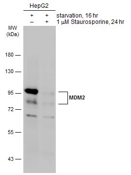
Untreated (–) and treated (+) HepG2 whole cell extracts (30 μg) were separated by 7.5% SDS-PAGE, and the membrane was blotted with MDM2 antibody (GTX70278) diluted at 1:1000. The HRP-conjugated anti-mouse IgG antibody (GTX213111-01) was used to detect the primary antibody, and the signal was developed with Trident ECL plus-Enhanced.
![IHC-P analysis of human breast carcinoma tissue using GTX70278 MDM2 antibody [SMP14].](https://www.genetex.com/upload/website/prouct_img/normal/GTX70278/GTX70278_4916_IHC-P_w_23061221_963.webp "IHC-P analysis of human breast carcinoma tissue using GTX70278 MDM2 antibody [SMP14].")
Untreated (–) and treated (+) HepG2 whole cell extracts (30 μg) were separated by 7.5% SDS-PAGE, and the membrane was blotted with MDM2 antibody (GTX70278) diluted at 1:1000. The HRP-conjugated anti-mouse IgG antibody (GTX213111-01) was used to detect the primary antibody, and the signal was developed with Trident ECL plus-Enhanced.
MDM2 antibody [SMP14]
GTX70278
ApplicationsImmunoPrecipitation, Western Blot, ImmunoHistoChemistry, ImmunoHistoChemistry Frozen, ImmunoHistoChemistry Paraffin
Product group Antibodies
ReactivityHuman, Mouse
TargetMDM2
Overview
- SupplierGeneTex
- Product NameMDM2 antibody [SMP14]
- Delivery Days Customer9
- Application Supplier NoteWB: 1:500-1:3000. IHC-P: 1/200-1/500. *Optimal dilutions/concentrations should be determined by the researcher.Not tested in other applications.
- ApplicationsImmunoPrecipitation, Western Blot, ImmunoHistoChemistry, ImmunoHistoChemistry Frozen, ImmunoHistoChemistry Paraffin
- CertificationResearch Use Only
- ClonalityMonoclonal
- Clone IDSMP14
- Concentration1 mg/ml
- ConjugateUnconjugated
- Gene ID4193
- Target nameMDM2
- Target descriptionMDM2 proto-oncogene
- Target synonymsACTFS, HDMX, LSKB, hdm2, E3 ubiquitin-protein ligase Mdm2, MDM2 oncogene, E3 ubiquitin protein ligase, MDM2 proto-oncogene, E3 ubiquitin protein ligase, Mdm2, p53 E3 ubiquitin protein ligase homolog, Mdm2, transformed 3T3 cell double minute 2, p53 binding protein, double minute 2, human homolog of; p53-binding protein, oncoprotein Mdm2
- HostMouse
- IsotypeIgG1
- Protein IDQ00987
- Protein NameE3 ubiquitin-protein ligase Mdm2
- Scientific DescriptionThis gene encodes a nuclear-localized E3 ubiquitin ligase. The encoded protein can promote tumor formation by targeting tumor suppressor proteins, such as p53, for proteasomal degradation. This gene is itself transcriptionally-regulated by p53. Overexpression or amplification of this locus is detected in a variety of different cancers. There is a pseudogene for this gene on chromosome 2. Alternative splicing results in a multitude of transcript variants, many of which may be expressed only in tumor cells. [provided by RefSeq, Jun 2013]
- ReactivityHuman, Mouse
- Storage Instruction-20°C or -80°C,2°C to 8°C
- UNSPSC41116161
Datasheet
Related products
Product group Antibodies
MDM2 AntibodyCSB-PA003212
ApplicationsImmunoFluorescence, Western Blot, ELISA, ImmunoHistoChemistry
ReactivityHuman, Monkey, Mouse
TargetMDM2
- SizePrice

Product group Antibodies
Anti-MDM2 Antibody Picoband(r)A00054-2-CARRIER-FREE
ApplicationsWestern Blot
ReactivityHuman, Mouse, Rat
TargetMDM2
- SizePrice
Product group Antibodies
Anti-MDM2 AntibodyA101381
ApplicationsWestern Blot, ELISA
ReactivityHuman
- SizePrice
Product group Antibodies
Anti-MDM2 Antibody144-60282
ApplicationsWestern Blot
ReactivityHuman, Mouse, Rat
TargetMDM2
- SizePrice
Product group Antibodies

Goat anti-MDM2 (isoform)EB06990
ApplicationsWestern Blot, ELISA
ReactivityBovine, Canine, Feline, Human, Porcine, Rat
TargetMDM2
- SizePrice
Product group Antibodies

MDM2 Antibody (C-Terminus)LS-C368433
ApplicationsWestern Blot
ReactivityBovine, Canine, Human, Mouse, Rat
TargetMDM2
- SizePrice

Product group Antibodies
ApplicationsFlow Cytometry, ImmunoFluorescence, Western Blot, ELISA, ImmunoCytoChemistry, ImmunoHistoChemistry, ImmunoHistoChemistry Frozen, ImmunoHistoChemistry Paraffin
ReactivityEquine, Human, Mouse, Rabbit, Rat
TargetMDM2
- SizePrice
![MDM2 antibody detects MDM2 protein at cytoplasm and nucleus by immunofluorescent analysis. Sample: HepG2 cells were fixed in 4% paraformaldehyde at RT for 15 min. Green: MDM2 stained by MDM2 antibody (GTX100531) diluted at 1:500. Red: alpha Tubulin, a cytoskeleton marker, stained by alpha Tubulin antibody [GT114] (GTX628802) diluted at 1:1000. Scale bar= 10μm.](https://www.genetex.com/upload/website/prouct_img/normal/GTX100531/GTX100531_44608_20220624_ICC_IF_22062919_841.webp)
Product group Antibodies
MDM2 antibodyGTX100531
ApplicationsImmunoFluorescence, ImmunoPrecipitation, Western Blot, ChIP Chromatin ImmunoPrecipitation, ImmunoCytoChemistry, ImmunoHistoChemistry, ImmunoHistoChemistry Paraffin
ReactivityHuman, Mouse
TargetMDM2
- SizePrice