

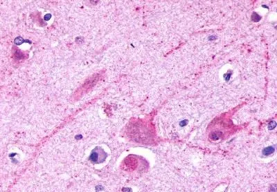
IHC-P analysis of brain, cortex neurons tissue using GTX13192 mGluR2 antibody.


IHC-P analysis of brain, cortex neurons tissue using GTX13192 mGluR2 antibody.
mGluR2 antibody
GTX13192
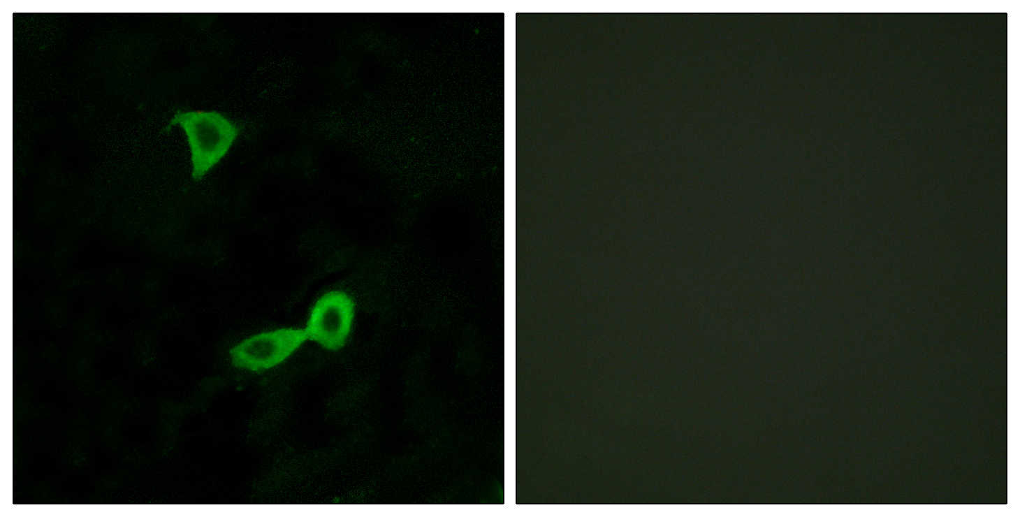
ApplicationsImmunoFluorescence, Western Blot, ImmunoCytoChemistry, ImmunoHistoChemistry, ImmunoHistoChemistry Paraffin
Product group Antibodies
ReactivityBovine, Canine, Hamster, Human, Mammals, Mouse, Porcine, Rabbit, Rat
TargetGRM2
Overview
- SupplierGeneTex
- Product NamemGluR2 antibody
- Delivery Days Customer9
- Application Supplier NoteIHC-P: 5 - 10 microg/ml. IHC-P: 5 - 10 microg/ml. *Optimal dilutions/concentrations should be determined by the researcher.Not tested in other applications.
- ApplicationsImmunoFluorescence, Western Blot, ImmunoCytoChemistry, ImmunoHistoChemistry, ImmunoHistoChemistry Paraffin
- CertificationResearch Use Only
- ClonalityPolyclonal
- Concentration1 mg/ml
- ConjugateUnconjugated
- Gene ID2912
- Target nameGRM2
- Target descriptionglutamate metabotropic receptor 2
- Target synonymsGLUR2, GPRC1B, MGLUR2, mGlu2, metabotropic glutamate receptor 2, glutamate receptor homolog, glutamate receptor, metabotropic 2
- HostRabbit
- IsotypeIgG
- Protein IDQ14416
- Protein NameMetabotropic glutamate receptor 2
- Scientific DescriptionL-glutamate is the major excitatory neurotransmitter in the central nervous system and activates both ionotropic and metabotropic glutamate receptors. Glutamatergic neurotransmission is involved in most aspects of normal brain function and can be perturbed in many neuropathologic conditions. The metabotropic glutamate receptors are a family of G protein-coupled receptors, that have been divided into 3 groups on the basis of sequence homology, putative signal transduction mechanisms, and pharmacologic properties. Group I includes GRM1 and GRM5 and these receptors have been shown to activate phospholipase C. Group II includes GRM2 and GRM3 while Group III includes GRM4, GRM6, GRM7 and GRM8. Group II and III receptors are linked to the inhibition of the cyclic AMP cascade but differ in their agonist selectivities. Several transcript variants encoding different isoforms have been found for this gene. [provided by RefSeq, Mar 2017]
- ReactivityBovine, Canine, Hamster, Human, Mammals, Mouse, Porcine, Rabbit, Rat
- Storage Instruction-20°C or -80°C,2°C to 8°C
- UNSPSC41116161
Datasheet
Related products
Product group Antibodies
GRM2 AntibodyCSB-PA009020
ApplicationsImmunoFluorescence, ELISA, ImmunoHistoChemistry
ReactivityHuman, Mouse, Rat
TargetGRM2
- SizePrice
Product group Antibodies
Anti-GRM2 AntibodyA96189
ApplicationsImmunoFluorescence, ELISA, ImmunoHistoChemistry
ReactivityHuman, Mouse, Rat
- SizePrice

Product group Antibodies
Anti-Metabotropic glutamate receptor 2/GRM2 Antibody Picoband(r)A06123-1-CARRIER-FREE
ApplicationsFlow Cytometry, Western Blot, ELISA, ImmunoHistoChemistry
ReactivityHuman, Mouse, Rat
TargetGRM2
- SizePrice
Product group Antibodies

GRM2 / MGLUR2 AntibodyLS-C831084
ApplicationsELISA, ImmunoHistoChemistry
ReactivityHuman, Mouse, Rat
TargetGRM2
- SizePrice
Product group Antibodies

Goat anti-GRM2EB10214
ApplicationsWestern Blot, ELISA, ImmunoHistoChemistry
ReactivityBovine, Canine, Human, Mouse, Rat
TargetGRM2
- SizePrice
Product group Antibodies
Anti-GRM2 AntibodyHPA065166
ApplicationsImmunoHistoChemistry
ReactivityHuman
TargetGRM2
- SizePrice
Product group Antibodies
GRM2 Polyclonal AntibodyCAC15129
ApplicationsImmunoFluorescence, Western Blot, ELISA
ReactivityRat
TargetGRM2
- SizePrice
Product group Antibodies
GRM2/GRM3 AntibodyPACO03993
ApplicationsWestern Blot, ELISA, ImmunoHistoChemistry
ReactivityHuman, Mouse, Rat
TargetGRM2
- SizePrice
Product group Antibodies
GRM2 Recombinant Antibody, Biotin ConjugatedBSM-61365R-BIOTIN
ApplicationsWestern Blot, ImmunoHistoChemistry, ImmunoHistoChemistry Frozen, ImmunoHistoChemistry Paraffin
ReactivityHuman, Mouse, Rat
TargetGRM2
- SizePrice