

Mitogen Activated Protein Kinase Kinase Kinase 7 Interacting Protein 1 (MAP3K7IP1) Polyclonal Antibody
CAU21420
ApplicationsImmunoPrecipitation, Western Blot, ImmunoCytoChemistry, ImmunoHistoChemistry
Product group Antibodies
ReactivityRat
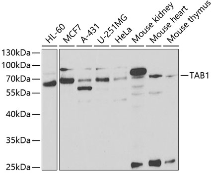
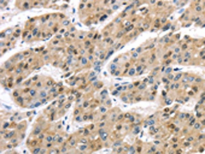
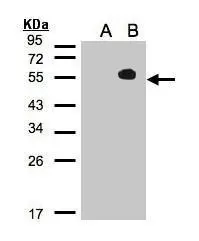
TargetTAB1
Overview
- SupplierBiomatik
- Product NameMitogen Activated Protein Kinase Kinase Kinase 7 Interacting Protein 1 (MAP3K7IP1) Polyclonal Antibody
- Delivery Days Customer12
- ApplicationsImmunoPrecipitation, Western Blot, ImmunoCytoChemistry, ImmunoHistoChemistry
- Applications SupplierWB; IHC; ICC; IP.
- CertificationResearch Use Only
- ClonalityPolyclonal
- Concentration0.5 mg/ml
- ConjugateUnconjugated
- Gene ID10454
- Target nameTAB1
- Target descriptionTGF-beta activated kinase 1 (MAP3K7) binding protein 1
- Target synonyms3'-Tab1, MAP3K7IP1, TGF-beta-activated kinase 1 and MAP3K7-binding protein 1, TAK1-binding protein 1, mitogen-activated protein kinase kinase kinase 7-interacting protein 1, transforming growth factor beta-activated kinase-binding protein 1
- HostRabbit
- Protein IDQ15750
- Protein NameTGF-beta-activated kinase 1 and MAP3K7-binding protein 1
- Scientific DescriptionThe Mitogen Activated Protein Kinase Kinase Kinase 7 Interacting Protein 1 (MAP3K7IP1) Polyclonal Antibody (Species: Human) has been validated for the following applications: WB, IHC, ICC, IP.
- ReactivityRat
- Reactivity SupplierHuman
- Storage Instruction-20°C,2°C to 8°C
- UNSPSC12352203
Related products

Product group Antibodies
Anti-TAB1 Antibody Picoband(r)A02847-1-CARRIER-FREE
ApplicationsWestern Blot, ELISA
ReactivityHuman
TargetTAB1
- SizePrice
Product group Antibodies
Anti-TAB1 Antibody144-05749
ApplicationsImmunoFluorescence, Western Blot
ReactivityHuman, Mouse
TargetTAB1
- SizePrice

Product group Antibodies
Anti-TAB1 AntibodyA14949
ApplicationsImmunoFluorescence, ImmunoPrecipitation, Western Blot, ImmunoCytoChemistry
ReactivityHuman, Mouse
- SizePrice
Product group Antibodies

TAB1 AntibodyLS-C814129
ApplicationsWestern Blot
ReactivityBovine, Canine, Human, Mouse, Porcine, Rat
TargetTAB1
- SizePrice
Product group Antibodies
TAB1 Recombinant AntibodyBSM-62019R
ApplicationsFlow Cytometry, ImmunoFluorescence, ImmunoPrecipitation, Western Blot, ImmunoCytoChemistry, ImmunoHistoChemistry, ImmunoHistoChemistry Frozen, ImmunoHistoChemistry Paraffin
ReactivityHuman, Mouse, Rat
TargetTAB1
- SizePrice
Product group Antibodies
TAB1 AntibodyCSB-PA251266
ApplicationsELISA, ImmunoHistoChemistry
ReactivityHuman, Mouse
TargetTAB1
- SizePrice
Product group Antibodies
TAB1 antibodyGTX103200
ApplicationsImmunoFluorescence, Western Blot, ImmunoCytoChemistry
ReactivityHuman
TargetTAB1
- SizePrice

Product group Antibodies
Anti-TAB1 AntibodyHPA039988
ApplicationsImmunoCytoChemistry, ImmunoHistoChemistry
ReactivityHuman
TargetTAB1
- SizePrice