

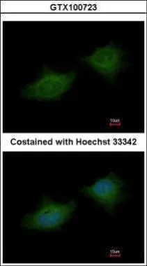
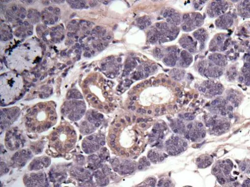
Immunofluorescence analysis of methanol-fixed HeLa, using MMP3(GTX100723) antibody at 1:200 dilution.
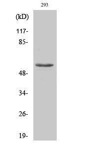
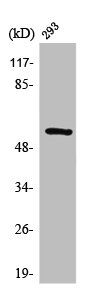
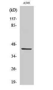
![Various whole cell extracts (30 μg) were separated by 10% SDS-PAGE, and the membrane was blotted with MMP3 antibody [N3C1], Internal (GTX100723) diluted at 1:1000. The HRP-conjugated anti-rabbit IgG antibody (GTX213110-01) was used to detect the primary antibody, and the signal was developed with Trident ECL plus-Enhanced.](https://www.genetex.com/upload/website/prouct_img/normal/GTX100723/GTX100723_39988_20210507_WB_w_23060100_331.webp "Various whole cell extracts (30 μg) were separated by 10% SDS-PAGE, and the membrane was blotted with MMP3 antibody [N3C1], Internal (GTX100723) diluted at 1:1000. The HRP-conjugated anti-rabbit IgG antibody (GTX213110-01) was used to detect the primary antibody, and the signal was developed with Trident ECL plus-Enhanced.")
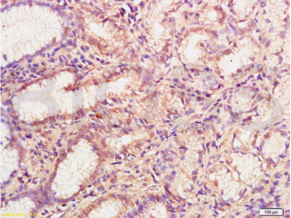
 antibody at 1:500 dilution.
Antigen Retrieval: Trilogy? (EDTA based, pH 8.0) buffer, 15min")
![Various whole cell extracts (30 μg) were separated by 10% SDS-PAGE, and the membrane was blotted with MMP3 antibody [N3C1], Internal (GTX100723) diluted at 1:1000. The HRP-conjugated anti-rabbit IgG antibody (GTX213110-01) was used to detect the primary antibody, and the signal was developed with Trident ECL plus-Enhanced.](https://www.genetex.com/upload/website/prouct_img/normal/GTX100723/GTX100723_39988_20210507_1_WB_w_23060100_742.webp "Various whole cell extracts (30 μg) were separated by 10% SDS-PAGE, and the membrane was blotted with MMP3 antibody [N3C1], Internal (GTX100723) diluted at 1:1000. The HRP-conjugated anti-rabbit IgG antibody (GTX213110-01) was used to detect the primary antibody, and the signal was developed with Trident ECL plus-Enhanced.")

Immunofluorescence analysis of methanol-fixed HeLa, using MMP3(GTX100723) antibody at 1:200 dilution.
MMP3 antibody [N3C1], Internal
GTX100723
ApplicationsImmunoFluorescence, Western Blot, ImmunoCytoChemistry, ImmunoHistoChemistry, ImmunoHistoChemistry Paraffin
Product group Antibodies
ReactivityHuman
TargetMMP3
Overview
- SupplierGeneTex
- Product NameMMP3 antibody [N3C1], Internal
- Delivery Days Customer9
- Application Supplier NoteWB: 1:500-1:3000. ICC/IF: 1:100-1:1000. IHC-P: 1:100-1:1000. *Optimal dilutions/concentrations should be determined by the researcher.Not tested in other applications.
- ApplicationsImmunoFluorescence, Western Blot, ImmunoCytoChemistry, ImmunoHistoChemistry, ImmunoHistoChemistry Paraffin
- CertificationResearch Use Only
- ClonalityPolyclonal
- Concentration1 mg/ml
- ConjugateUnconjugated
- Gene ID4314
- Target nameMMP3
- Target descriptionmatrix metallopeptidase 3
- Target synonymsCHDS6, MMP-3, SL-1, STMY, STMY1, STR1, stromelysin-1, matrix metalloproteinase 3 (stromelysin 1, progelatinase), proteoglycanase, transin-1
- HostRabbit
- IsotypeIgG
- Protein IDP08254
- Protein NameStromelysin-1
- Scientific DescriptionProteins of the matrix metalloproteinase (MMP) family are involved in the breakdown of extracellular matrix in normal physiological processes, such as embryonic development, reproduction, and tissue remodeling, as well as in disease processes, such as arthritis and metastasis. Most MMPs are secreted as inactive proproteins which are activated when cleaved by extracellular proteinases. This gene encodes an enzyme which degrades fibronectin, laminin, collagens III, IV, IX, and X, and cartilage proteoglycans. The enzyme is thought to be involved in wound repair, progression of atherosclerosis, and tumor initiation. The gene is part of a cluster of MMP genes which localize to chromosome 11q22.3. [provided by RefSeq]
- ReactivityHuman
- Storage Instruction-20°C or -80°C,2°C to 8°C
- UNSPSC41116161
Datasheet
Related products
Product group Antibodies
Anti-MMP3 Antibody144-01202
ApplicationsWestern Blot
ReactivityHuman, Mouse
TargetMMP3
- SizePrice
Product group Antibodies
Anti-MMP-3 AntibodyA34461
ApplicationsImmunoFluorescence, Western Blot, ELISA, ImmunoHistoChemistry
ReactivityHuman, Mouse, Rat
- SizePrice
Product group Antibodies
References
MMP3 Polyclonal AntibodyBS-0413R
ApplicationsImmunoFluorescence, Western Blot, ELISA, ImmunoCytoChemistry, ImmunoHistoChemistry, ImmunoHistoChemistry Frozen, ImmunoHistoChemistry Paraffin
ReactivityHuman
TargetMMP3
- SizePrice
Product group Antibodies
MMP3 AntibodyCSB-PA004191
ApplicationsImmunoFluorescence, Western Blot, ELISA, ImmunoHistoChemistry
ReactivityHuman, Mouse, Rat
TargetMMP3
- SizePrice
Product group Antibodies

Goat anti-MMP3 (aa459-473)EB11719
ApplicationsWestern Blot, ELISA, ImmunoHistoChemistry
ReactivityHuman, Mouse
TargetMMP3
- SizePrice
Product group Antibodies
Mmp3 Polyclonal AntibodyCAC07007
ApplicationsWestern Blot, ELISA, ImmunoHistoChemistry
ReactivityMouse
TargetMMP3
- SizePrice
![IHC-P analysis of human spleen tissue section using GTX02675 MMP3 antibody [rMMP3/1730].](https://www.genetex.com/upload/website/prouct_img/normal/GTX02675/GTX02675_20210319_IHC-P_w_23053122_889.webp)
Product group Antibodies
MMP3 antibody [rMMP3/1730]GTX02675
ApplicationsImmunoHistoChemistry, ImmunoHistoChemistry Paraffin
ReactivityHuman
TargetMMP3
- SizePrice
Product group Antibodies
MMP3 antibody [N1], N-termGTX100706
ApplicationsImmunoFluorescence, ImmunoCytoChemistry, ImmunoHistoChemistry, ImmunoHistoChemistry Paraffin
ReactivityHuman
TargetMMP3
- SizePrice
Product group Antibodies
MMP3 (cleaved Phe100) antibodyGTX86941
ApplicationsWestern Blot
ReactivityHuman
TargetMMP3
- SizePrice