

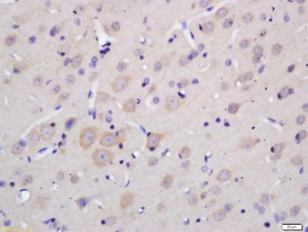
IHC-P analysis of rat brain tissue using GTX03460 MOBP antibody. Dilution : 1:200
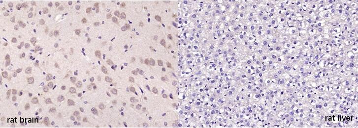
 and rat liver (right) tissue using GTX03460 MOBP antibody. Antigen retrieval : Boiling in sodium citrate buffer (pH6.0) for 15min Dilution : 1:200")

IHC-P analysis of rat brain tissue using GTX03460 MOBP antibody. Dilution : 1:200
MOBP antibody
GTX03460
ApplicationsWestern Blot, ImmunoHistoChemistry, ImmunoHistoChemistry Paraffin
Product group Antibodies
ReactivityHuman, Mouse, Rat
TargetMOBP
Overview
- SupplierGeneTex
- Product NameMOBP antibody
- Delivery Days Customer9
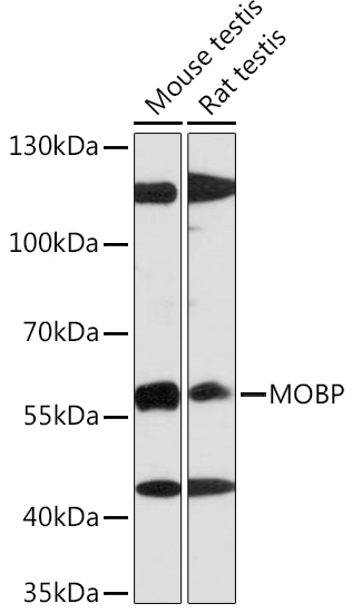
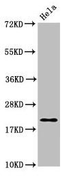
- Application Supplier NoteWB: 1:500-1:2000. IHC-P: 1:100-1:500. *Optimal dilutions/concentrations should be determined by the researcher.Not tested in other applications.
- ApplicationsWestern Blot, ImmunoHistoChemistry, ImmunoHistoChemistry Paraffin
- CertificationResearch Use Only
- ClonalityPolyclonal
- Concentration1 mg/ml
- ConjugateUnconjugated
- Gene ID4336
- Target nameMOBP
- Target descriptionmyelin associated oligodendrocyte basic protein
- Target synonymsmyelin-associated oligodendrocyte basic protein
- HostRabbit
- IsotypeIgG
- Protein IDQ13875
- Protein NameMyelin-associated oligodendrocyte basic protein
- Scientific DescriptionPredicted to enable actin binding activity and myosin binding activity. Predicted to be a structural constituent of myelin sheath. Predicted to be involved in nervous system development. Predicted to be located in mitochondrion. Predicted to be active in cortical actin cytoskeleton. Implicated in frontotemporal dementia. [provided by Alliance of Genome Resources, Nov 2021]
- ReactivityHuman, Mouse, Rat
- Storage Instruction-20°C or -80°C,2°C to 8°C
- UNSPSC12352203
Datasheet
Related products
Product group Antibodies
Anti-MOBP AntibodyA91716
ApplicationsImmunoFluorescence, Western Blot, ImmunoCytoChemistry
ReactivityHuman, Mouse, Rat
- SizePrice
Product group Antibodies
MOBP Polyclonal AntibodyCAC13118
ApplicationsImmunoFluorescence, Western Blot, ELISA
TargetMOBP
- SizePrice
Product group Antibodies
References
MOBP Polyclonal AntibodyBS-11184R
ApplicationsImmunoFluorescence, Western Blot, ELISA, ImmunoCytoChemistry, ImmunoHistoChemistry, ImmunoHistoChemistry Frozen, ImmunoHistoChemistry Paraffin
TargetMOBP
- SizePrice
Product group Antibodies
Anti-MOBP Antibody144-64901
ApplicationsWestern Blot
ReactivityHuman, Mouse, Rat
TargetMOBP
- SizePrice
Product group Antibodies

MOBP AntibodyLS-C670686
ApplicationsImmunoFluorescence, Western Blot, ELISA
ReactivityHuman
TargetMOBP
- SizePrice

Product group Antibodies
Anti-MOBP AntibodyHPA035152
ApplicationsImmunoHistoChemistry
ReactivityHuman
TargetMOBP
- SizePrice
Product group Antibodies
MOBP AntibodyCSB-PA014701LA01HU
ApplicationsImmunoFluorescence, Western Blot, ELISA
ReactivityHuman
TargetMOBP
- SizePrice

Product group Antibodies
Anti-MOBP Antibody Picoband(r)A09516-2-CARRIER-FREE
ApplicationsFlow Cytometry, Western Blot, ELISA
ReactivityHuman, Mouse, Rat
TargetMOBP
- SizePrice