

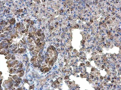
Monoamine Oxidase B antibody [N2C3] detects Monoamine Oxidase B protein at cytoplasm on mouse lung by immunohistochemical analysis. Sample: Paraffin-embedded mouse lung. Monoamine Oxidase B antibody [N2C3] (GTX105970) diluted at 1:500.
Antigen Retrieval: Trilogy? (EDTA based, pH 8.0) buffer, 15min
![Monoamine Oxidase B antibody [N2C3] detects Monoamine Oxidase B protein at cytoplasm by immunohistochemical analysis. Sample: Paraffin-embedded mouse liver. Monoamine Oxidase B stained by Monoamine Oxidase B antibody [N2C3] (GTX105970) diluted at 1:500. Antigen Retrieval: Citrate buffer, pH 6.0, 15 min](https://www.genetex.com/upload/website/prouct_img/normal/GTX105970/GTX105970_43300_20190517_IHC-P_M_w_23060120_810.webp "Monoamine Oxidase B antibody [N2C3] detects Monoamine Oxidase B protein at cytoplasm by immunohistochemical analysis. Sample: Paraffin-embedded mouse liver. Monoamine Oxidase B stained by Monoamine Oxidase B antibody [N2C3] (GTX105970) diluted at 1:500. Antigen Retrieval: Citrate buffer, pH 6.0, 15 min")

![Monoamine Oxidase B antibody [N2C3] detects Monoamine Oxidase B protein at mitochondria by immunofluorescent analysis. Sample: HepG2 cells were fixed in ice-cold MeOH for 5 min. Green: Monoamine Oxidase B stained by Monoamine Oxidase B antibody [N2C3] (GTX105970) diluted at 1:500. Blue: Hoechst 33342 staining.](https://www.genetex.com/upload/website/prouct_img/normal/GTX105970/GTX105970_43300_20181205_ICC_IF_w_23060120_539.webp "Monoamine Oxidase B antibody [N2C3] detects Monoamine Oxidase B protein at mitochondria by immunofluorescent analysis. Sample: HepG2 cells were fixed in ice-cold MeOH for 5 min. Green: Monoamine Oxidase B stained by Monoamine Oxidase B antibody [N2C3] (GTX105970) diluted at 1:500. Blue: Hoechst 33342 staining.")
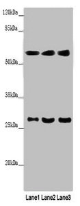
![Mouse tissue extract (50 μg) was separated by 7.5% SDS-PAGE, and the membrane was blotted with Monoamine Oxidase B antibody [N2C3] (GTX105970) diluted at 1:1000. The HRP-conjugated anti-rabbit IgG antibody (GTX213110-01) was used to detect the primary antibody.](https://www.genetex.com/upload/website/prouct_img/normal/GTX105970/GTX105970_43300_20230721_WB_M_liver_23072519_379.webp "Mouse tissue extract (50 μg) was separated by 7.5% SDS-PAGE, and the membrane was blotted with Monoamine Oxidase B antibody [N2C3] (GTX105970) diluted at 1:1000. The HRP-conjugated anti-rabbit IgG antibody (GTX213110-01) was used to detect the primary antibody.")
![HepG2 whole cell and mitochondria extracts (8.1 μg) were separated by 10% SDS-PAGE, and the membrane was blotted with Monoamine Oxidase B antibody [N2C3] (GTX105970) diluted at 1:3000. The HRP-conjugated anti-rabbit IgG antibody (GTX213110-01) was used to detect the primary antibody.](https://www.genetex.com/upload/website/prouct_img/normal/GTX105970/GTX105970_45196_20240216_WB_Fraction_24032600_876.webp "HepG2 whole cell and mitochondria extracts (8.1 μg) were separated by 10% SDS-PAGE, and the membrane was blotted with Monoamine Oxidase B antibody [N2C3] (GTX105970) diluted at 1:3000. The HRP-conjugated anti-rabbit IgG antibody (GTX213110-01) was used to detect the primary antibody.")

Monoamine Oxidase B antibody [N2C3] detects Monoamine Oxidase B protein at cytoplasm on mouse lung by immunohistochemical analysis. Sample: Paraffin-embedded mouse lung. Monoamine Oxidase B antibody [N2C3] (GTX105970) diluted at 1:500.
Antigen Retrieval: Trilogy? (EDTA based, pH 8.0) buffer, 15min
Monoamine Oxidase B antibody [N2C3]
GTX105970
ApplicationsImmunoFluorescence, Western Blot, ELISA, ImmunoCytoChemistry, ImmunoHistoChemistry, ImmunoHistoChemistry Paraffin
Product group Antibodies
ReactivityHuman, Mouse
TargetMAOB
Overview
- SupplierGeneTex
- Product NameMonoamine Oxidase B antibody [N2C3]
- Delivery Days Customer9
- Application Supplier NoteWB: 1:500-1:3000. ICC/IF: 1:100-1:1000. IHC-P: 1:100-1:1000. ELISA: 1:1000-1:10000. *Optimal dilutions/concentrations should be determined by the researcher.Not tested in other applications.
- ApplicationsImmunoFluorescence, Western Blot, ELISA, ImmunoCytoChemistry, ImmunoHistoChemistry, ImmunoHistoChemistry Paraffin
- CertificationResearch Use Only
- ClonalityPolyclonal
- Concentration0.51 mg/ml
- ConjugateUnconjugated
- Gene ID4129
- Target nameMAOB
- Target descriptionmonoamine oxidase B
- Target synonymsamine oxidase [flavin-containing] B, MAO, brain, MAO, platelet, MAO-B, adrenalin oxidase, monoamine oxidase type B, tyramine oxidase
- HostRabbit
- IsotypeIgG
- Protein IDP27338
- Protein NameAmine oxidase [flavin-containing] B
- Scientific DescriptionThe protein encoded by this gene belongs to the flavin monoamine oxidase family. It is a enzyme located in the mitochondrial outer membrane. It catalyzes the oxidative deamination of biogenic and xenobiotic amines and plays an important role in the metabolism of neuroactive and vasoactive amines in the central nervous sysytem and peripheral tissues. This protein preferentially degrades benzylamine and phenylethylamine. [provided by RefSeq]
- ReactivityHuman, Mouse
- Storage Instruction-20°C or -80°C,2°C to 8°C
- UNSPSC41116161
Datasheet
Related products
Product group Antibodies
ReactivityHuman
TargetMAOB
- SizePrice
Product group Antibodies
MAOB AntibodyCSB-PA013399LA01HU
ApplicationsWestern Blot, ELISA, ImmunoHistoChemistry
ReactivityHuman, Mouse
TargetMAOB
- SizePrice
Product group Antibodies

Monoamine Oxidase B (MAOB) AntibodyABX109527
ApplicationsWestern Blot, ELISA, ImmunoHistoChemistry
- SizePrice
Product group Antibodies
Anti-MAOB AntibodyA35480
ApplicationsImmunoFluorescence, Western Blot, ImmunoHistoChemistry
ReactivityHuman
- SizePrice
Product group Antibodies

ApplicationsWestern Blot, ELISA, ImmunoHistoChemistry, ImmunoHistoChemistry Paraffin
ReactivityHuman
TargetMAOB
- SizePrice
Product group Antibodies

Goat anti-MAOBEB06984
ApplicationsFlow Cytometry, ImmunoFluorescence, Western Blot, ELISA
ReactivityBovine, Canine, Human, Mouse, Porcine, Rat
TargetMAOB
- SizePrice

Product group Antibodies
Anti-MAOB-25ulHPA002328
ApplicationsWestern Blot, ImmunoHistoChemistry
ReactivityHuman, Mouse, Rat
- SizePrice
Product group Antibodies
MAOB Polyclonal AntibodyCAC13813
ApplicationsWestern Blot, ELISA, ImmunoHistoChemistry
ReactivityMouse
TargetMAOB
- SizePrice

Product group Antibodies
Anti-Monoamine Oxidase B/MAOB Antibody Picoband(r)PB9665-CARRIER-FREE
ApplicationsWestern Blot, ImmunoHistoChemistry
ReactivityHuman, Mouse, Rat
TargetMAOB
- SizePrice
![Rat tissue extract (50 μg) was separated by 10% SDS-PAGE, and the membrane was blotted with Monoamine Oxidase B antibody [N1N3] (GTX113771) diluted at 1:3000.](https://www.genetex.com/upload/website/prouct_img/normal/GTX113771/GTX113771_40142_20151105_WB_R_liver_w_23060501_168.webp)
Product group Antibodies
Monoamine Oxidase B antibody [N1N3]GTX113771
ApplicationsImmunoFluorescence, Western Blot, ImmunoCytoChemistry, ImmunoHistoChemistry, ImmunoHistoChemistry Paraffin
ReactivityHuman, Mouse, Rat
TargetMAOB
- SizePrice