

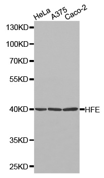
MSL2 Antibody
CAB13103
ReactivityHuman
Product group Antibodies
TargetHFE
Overview
- SupplierAssay Genie
- Product NameMSL2 Antibody
- Delivery Days Customer9
- Applications SupplierWB
- CertificationResearch Use Only
- ClonalityPolyclonal
- Gene ID3077
- Target nameHFE
- Target descriptionhomeostatic iron regulator
- Target synonymsHFE1, HH, HLA-H, MVCD7, TFQTL2, hereditary hemochromatosis protein, MHC class I-like protein HFE, hereditary hemochromatosis protein HLA-H, high Fe
- HostRabbit
- Protein IDQ30201
- Protein NameHereditary hemochromatosis protein
- Shelf life instruction12 months
- SourceRabbit
- ReactivityHuman
- Reactivity SupplierHuman,Mouse,Rat
- Storage Instruction-20°C
- UNSPSC12352203
Related products
Product group Antibodies
HFE AntibodyCSB-PA653744LA01HU
ApplicationsELISA
ReactivityHuman
TargetHFE
- SizePrice

Product group Antibodies
Anti-HFE Antibody Picoband(r)A00506-1-CARRIER-FREE
ApplicationsWestern Blot
ReactivityHuman
TargetHFE
- SizePrice
Product group Antibodies
Anti-HFE Antibody144-01310
ApplicationsImmunoFluorescence, Western Blot
ReactivityHuman, Mouse
TargetHFE
- SizePrice
Product group Antibodies
Anti-HFE AntibodyA29875
ApplicationsImmunoFluorescence, Western Blot, ImmunoHistoChemistry
ReactivityHuman
- SizePrice

Product group Antibodies
Anti-HFE AntibodyHPA017276
ApplicationsWestern Blot, ImmunoHistoChemistry
ReactivityHuman
TargetHFE
- SizePrice
Product group Antibodies

HFE AntibodyLS-C331410
ApplicationsImmunoFluorescence, Western Blot, ImmunoHistoChemistry
ReactivityHuman, Mouse
TargetHFE
- SizePrice
Product group Antibodies
References
HFE Polyclonal AntibodyBS-12335R
ApplicationsFlow Cytometry, ImmunoFluorescence, Western Blot, ELISA, ImmunoCytoChemistry, ImmunoHistoChemistry, ImmunoHistoChemistry Frozen, ImmunoHistoChemistry Paraffin
ReactivityBovine, Canine, Equine, Human, Mouse, Rat, Sheep
TargetHFE
- SizePrice
Product group Antibodies
HFE antibodyGTX135988
ApplicationsWestern Blot, ImmunoHistoChemistry, ImmunoHistoChemistry Paraffin
ReactivityHuman
TargetHFE
- SizePrice