

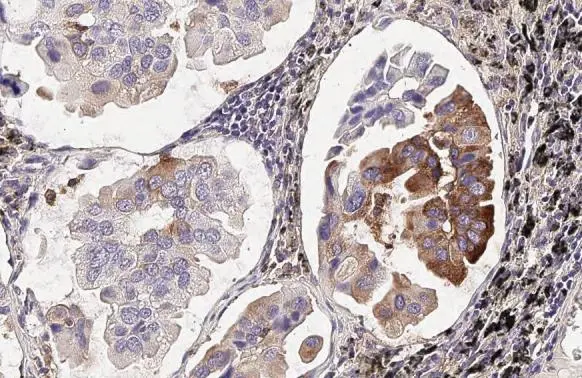
MX1 antibody [HL2051] detects MX1 protein at cytoplasm and nucleus by immunohistochemical analysis. Sample: Paraffin-embedded human lung cancer. MX1 stained by MX1 antibody [HL2051] (GTX637955) diluted at 1:100. Antigen Retrieval: Citrate buffer, pH 6.0, 15 min
![MX1 antibody [HL2051] detects MX1 protein at cytoplasm and nucleus by immunohistochemical analysis. Sample: Paraffin-embedded mouse kidney. MX1 stained by MX1 antibody [HL2051] (GTX637955) diluted at 1:100. Antigen Retrieval: Citrate buffer, pH 6.0, 15 min](https://www.genetex.com/upload/website/prouct_img/normal/GTX637955/GTX637955_T-44886_20221230_IHC-P_M_23020621_453.webp "MX1 antibody [HL2051] detects MX1 protein at cytoplasm and nucleus by immunohistochemical analysis. Sample: Paraffin-embedded mouse kidney. MX1 stained by MX1 antibody [HL2051] (GTX637955) diluted at 1:100. Antigen Retrieval: Citrate buffer, pH 6.0, 15 min")
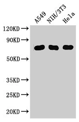
![Various whole cell extracts (30 μg) were separated by 7.5% SDS-PAGE, and the membrane was blotted with MX1 antibody [HL2051] (GTX637955) diluted at 1:1000. The HRP-conjugated anti-rabbit IgG antibody (GTX213110-01) was used to detect the primary antibody. Corresponding RNA expression data for the same cell lines are based on Human Protein Atlas program.](https://www.genetex.com/upload/website/prouct_img/normal/GTX637955/GTX637955_44963_20230224_WB_TPM_watermark_23030219_269.webp "Various whole cell extracts (30 μg) were separated by 7.5% SDS-PAGE, and the membrane was blotted with MX1 antibody [HL2051] (GTX637955) diluted at 1:1000. The HRP-conjugated anti-rabbit IgG antibody (GTX213110-01) was used to detect the primary antibody. Corresponding RNA expression data for the same cell lines are based on Human Protein Atlas program.")
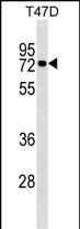
![Non-transfected (–) and transfected (+) 293T whole cell extracts (30 μg) were separated by 7.5% SDS-PAGE, and the membrane was blotted with MX1 antibody [HL2051] (GTX637955) diluted at 1:5000. The HRP-conjugated anti-rabbit IgG antibody (GTX213110-01) was used to detect the primary antibody.](https://www.genetex.com/upload/website/prouct_img/normal/GTX637955/GTX637955_44963_20230512_WB_multiple_B_23051702_696.webp "Non-transfected (–) and transfected (+) 293T whole cell extracts (30 μg) were separated by 7.5% SDS-PAGE, and the membrane was blotted with MX1 antibody [HL2051] (GTX637955) diluted at 1:5000. The HRP-conjugated anti-rabbit IgG antibody (GTX213110-01) was used to detect the primary antibody.")
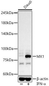
![Whole cell extract (30 μg) was separated by 7.5% SDS-PAGE, and the membrane was blotted with MX1 antibody [HL2051] (GTX637955) diluted at 1:1000. The HRP-conjugated anti-rabbit IgG antibody (GTX213110-01) was used to detect the primary antibody.](https://www.genetex.com/upload/website/prouct_img/normal/GTX637955/GTX637955_44963_20230616_WB_D_23062019_720.webp "Whole cell extract (30 μg) was separated by 7.5% SDS-PAGE, and the membrane was blotted with MX1 antibody [HL2051] (GTX637955) diluted at 1:1000. The HRP-conjugated anti-rabbit IgG antibody (GTX213110-01) was used to detect the primary antibody.")
![MX1 antibody [HL2051] detects MX1 protein at cytoplasm and nucleus by immunohistochemical analysis. Sample: Paraffin-embedded cat kidney. MX1 stained by MX1 antibody [HL2051] (GTX637955) diluted at 1:100. Antigen Retrieval: Citrate buffer, pH 6.0, 15 min](https://www.genetex.com/upload/website/prouct_img/normal/GTX637955/GTX637955_44963_20230721_IHC-P_Cat_23073119_720.webp "MX1 antibody [HL2051] detects MX1 protein at cytoplasm and nucleus by immunohistochemical analysis. Sample: Paraffin-embedded cat kidney. MX1 stained by MX1 antibody [HL2051] (GTX637955) diluted at 1:100. Antigen Retrieval: Citrate buffer, pH 6.0, 15 min")
![MX1 antibody [HL2051] detects MX1 protein by immunofluorescent analysis. Sample: MDCK cells were fixed in 4% paraformaldehyde at RT for 15 min. Green: MX1 stained by MX1 antibody [HL2051] (GTX637955) diluted at 1:500. Red: alpha Tubulin, a cytoskeleton marker, stained by alpha Tubulin antibody [GT114] (GTX628802) diluted at 1:1000. Blue: Fluoroshield with DAPI (GTX30920).](https://www.genetex.com/upload/website/prouct_img/normal/GTX637955/GTX637955_44963_20240726_ICC_IF_D_24081300_919.webp "MX1 antibody [HL2051] detects MX1 protein by immunofluorescent analysis. Sample: MDCK cells were fixed in 4% paraformaldehyde at RT for 15 min. Green: MX1 stained by MX1 antibody [HL2051] (GTX637955) diluted at 1:500. Red: alpha Tubulin, a cytoskeleton marker, stained by alpha Tubulin antibody [GT114] (GTX628802) diluted at 1:1000. Blue: Fluoroshield with DAPI (GTX30920).")
MX1 antibody [HL2051] detects MX1 protein at cytoplasm and nucleus by immunohistochemical analysis. Sample: Paraffin-embedded human lung cancer. MX1 stained by MX1 antibody [HL2051] (GTX637955) diluted at 1:100. Antigen Retrieval: Citrate buffer, pH 6.0, 15 min
MX1 antibody [HL2051]
GTX637955
ApplicationsImmunoFluorescence, Western Blot, ImmunoCytoChemistry, ImmunoHistoChemistry, ImmunoHistoChemistry Paraffin
Product group Antibodies
ReactivityCanine, Feline, Human, Mouse
TargetMX1
Overview
- SupplierGeneTex
- Product NameMX1 antibody [HL2051]
- Delivery Days Customer9
- Application Supplier NoteWB: 1:500-1:3000. *Optimal dilutions/concentrations should be determined by the researcher.Not tested in other applications.
- ApplicationsImmunoFluorescence, Western Blot, ImmunoCytoChemistry, ImmunoHistoChemistry, ImmunoHistoChemistry Paraffin
- CertificationResearch Use Only
- ClonalityMonoclonal
- Clone IDHL2051
- Concentration1 mg/ml
- ConjugateUnconjugated
- Gene ID4599
- Target nameMX1
- Target descriptionMX dynamin like GTPase 1
- Target synonymsIFI-78K, IFI78, MX, MxA, lncMX1-215, interferon-induced GTP-binding protein Mx1, interferon-induced protein p78, interferon-inducible protein p78, interferon-regulated resistance GTP-binding protein MxA, myxoma resistance protein 1, myxovirus (influenza virus) resistance 1, interferon-inducible protein p78
- HostRabbit
- IsotypeIgG
- Protein IDP20591
- Protein NameInterferon-induced GTP-binding protein Mx1
- Scientific DescriptionThis gene encodes a guanosine triphosphate (GTP)-metabolizing protein that participates in the cellular antiviral response. The encoded protein is induced by type I and type II interferons and antagonizes the replication process of several different RNA and DNA viruses. There is a related gene located adjacent to this gene on chromosome 21, and there are multiple pseudogenes located in a cluster on chromosome 4. Alternative splicing results in multiple transcript variants. [provided by RefSeq, Sep 2013]
- ReactivityCanine, Feline, Human, Mouse
- Storage Instruction-20°C or -80°C,2°C to 8°C
- UNSPSC41116161
Datasheet
Related products
Product group Antibodies
MX1 AntibodyCSB-PA015249LA01HU
ApplicationsImmunoFluorescence, ImmunoPrecipitation, Western Blot, ELISA, ImmunoHistoChemistry
ReactivityHuman, Mouse
TargetMX1
- SizePrice

Product group Antibodies
Anti-MX1 Antibody Picoband(r)A00849-1-CARRIER-FREE
ApplicationsFlow Cytometry, Western Blot, ELISA
ReactivityHuman, Mouse
TargetMX1
- SizePrice
Product group Antibodies
Anti-MX1 AntibodyA11481
ApplicationsImmunoFluorescence, Western Blot, ImmunoCytoChemistry, ImmunoHistoChemistry
ReactivityHuman, Mouse
- SizePrice
Product group Antibodies
Anti-MX1 Antibody144-65170
ApplicationsWestern Blot
ReactivityHuman, Mouse
TargetMX1
- SizePrice

Product group Antibodies
Anti-MX1 AntibodyHPA030917
ApplicationsWestern Blot, ImmunoHistoChemistry
ReactivityHuman
TargetMX1
- SizePrice
Product group Antibodies

MX1 / MX AntibodyLS-C331685
ApplicationsWestern Blot
ReactivityHuman, Mouse
TargetMX1
- SizePrice
Product group Antibodies
MX1 Monoclonal AntibodyBSM-51528M
ApplicationsWestern Blot
ReactivityHuman, Porcine
TargetMX1
- SizePrice
![Non-transfected (–) and transfected (+) 293T whole cell extracts (30 μg) were separated by 7.5% SDS-PAGE, and the membrane was blotted with MX1 antibody [HL1790] (GTX637442) diluted at 1:5000. The HRP-conjugated anti-rabbit IgG antibody (GTX213110-01) was used to detect the primary antibody.](https://www.genetex.com/upload/website/prouct_img/normal/GTX637442/GTX637442_T-44809_20221223_WB_multiple_B_22122722_968.webp)
Product group Antibodies
MX1 antibody [HL1790]GTX637442
ApplicationsImmunoFluorescence, Western Blot, ImmunoCytoChemistry, ImmunoHistoChemistry, ImmunoHistoChemistry Paraffin
ReactivityFeline, Human
TargetMX1
- SizePrice
![IHC-P analysis of human laryngeal squamous cell carcinoma (LSCC) tissue using GTX639940 MX1 antibody [HMV316] HistoMAX?. Squamous cell carcinoma with strong MX1 staining of tumor cells. Stromal and inflammatory cells show a less intense MX1 staining.](https://www.genetex.com/upload/website/prouct_img/normal/GTX639940/GTX639940_20240408_IHC-P_24040801_462.webp)
Product group Antibodies
MX1 antibody [HMV316] HistoMAX(tm)GTX639940
ApplicationsImmunoHistoChemistry, ImmunoHistoChemistry Paraffin
ReactivityHuman
TargetMX1
- SizePrice