

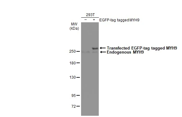
Non-transfected (–) and transfected (+) 293T whole cell extracts (30 μg) were separated by 5% SDS-PAGE, and the membrane was blotted with MYH9 antibody [N1], N-term (GTX101751) diluted at 1:5000. The HRP-conjugated anti-rabbit IgG antibody (GTX213110-01) was used to detect the primary antibody.
![Various whole cell extracts (30 μg) were separated by 5% SDS-PAGE, and the membrane was blotted with MYH9 antibody [N1], N-term (GTX101751) diluted at 1:1000. The HRP-conjugated anti-rabbit IgG antibody (GTX213110-01) was used to detect the primary antibody.](https://www.genetex.com/upload/website/prouct_img/normal/GTX101751/GTX101751_43628_20190628_WB_22081423_799.webp "Various whole cell extracts (30 μg) were separated by 5% SDS-PAGE, and the membrane was blotted with MYH9 antibody [N1], N-term (GTX101751) diluted at 1:1000. The HRP-conjugated anti-rabbit IgG antibody (GTX213110-01) was used to detect the primary antibody.")
![Immunoprecipitation of MYH9 protein from HeLa whole cell extracts using 5 μg of MYH9 antibody [N1], N-term (GTX101751). Western blot analysis was performed using MYH9 antibody [N1], N-term (GTX101751). EasyBlot anti-Rabbit IgG (GTX221666-01) was used as a secondary reagent.](https://www.genetex.com/upload/website/prouct_img/normal/GTX101751/GTX101751_40632_20150508_IP_w_23060100_841.webp "Immunoprecipitation of MYH9 protein from HeLa whole cell extracts using 5 μg of MYH9 antibody [N1], N-term (GTX101751). Western blot analysis was performed using MYH9 antibody [N1], N-term (GTX101751). EasyBlot anti-Rabbit IgG (GTX221666-01) was used as a secondary reagent.")
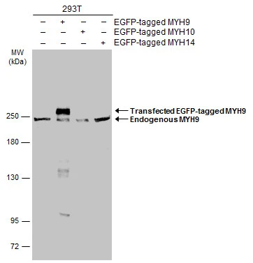
![Non-transfected (–) and transfected (+) 293T whole cell extracts (30 μg) were separated by 5% SDS-PAGE, and the membrane was blotted with MYH9 antibody [N1], N-term (GTX101751) diluted at 1:2000. The HRP-conjugated anti-rabbit IgG antibody (GTX213110-01) was used to detect the primary antibody.](https://www.genetex.com/upload/website/prouct_img/normal/GTX101751/GTX101751_40632_20191129_WB_shRNA_watermark_w_23060100_955.webp "Non-transfected (–) and transfected (+) 293T whole cell extracts (30 μg) were separated by 5% SDS-PAGE, and the membrane was blotted with MYH9 antibody [N1], N-term (GTX101751) diluted at 1:2000. The HRP-conjugated anti-rabbit IgG antibody (GTX213110-01) was used to detect the primary antibody.")
![MYH9 antibody [N1], N-term detects MYH9 protein by Western blot analysis. A. 30 μg U87-MG whole cell lysate/extract B. 30 μg SK-N-SH whole cell lysate/extract C. 30 μg IMR32 whole cell lysate/extract D. 30 μg SK-N-AS whole cell lysate/extract 5 % SDS-PAGE MYH9 antibody [N1], N-term (GTX101751) dilution: 1:1000](https://www.genetex.com/upload/website/prouct_img/normal/GTX101751/GTX101751_40632_WB_1_w_23060100_229.webp "MYH9 antibody [N1], N-term detects MYH9 protein by Western blot analysis. A. 30 μg U87-MG whole cell lysate/extract B. 30 μg SK-N-SH whole cell lysate/extract C. 30 μg IMR32 whole cell lysate/extract D. 30 μg SK-N-AS whole cell lysate/extract 5 % SDS-PAGE MYH9 antibody [N1], N-term (GTX101751) dilution: 1:1000")
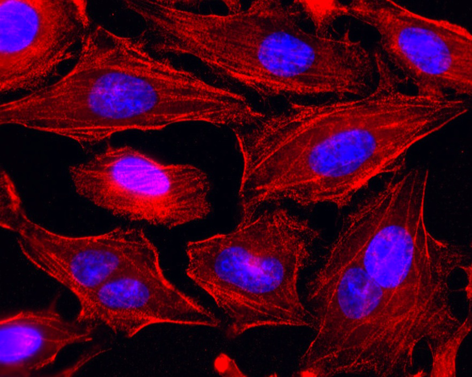
![MYH9 antibody [N1], N-term detects MYH9 protein at cytoskeleton by immunofluorescent analysis. Sample: HeLa cells were fixed in ice-cold MeOH for 5 min. Green: MYH9 stained by MYH9 antibody [N1], N-term (GTX101751) diluted at 1:500. Blue: Fluoroshield with DAPI (GTX30920). Scale bar= 10 μm.](https://www.genetex.com/upload/website/prouct_img/normal/GTX101751/GTX101751_43628_20200219_ICC_IF_w_23060100_596.webp "MYH9 antibody [N1], N-term detects MYH9 protein at cytoskeleton by immunofluorescent analysis. Sample: HeLa cells were fixed in ice-cold MeOH for 5 min. Green: MYH9 stained by MYH9 antibody [N1], N-term (GTX101751) diluted at 1:500. Blue: Fluoroshield with DAPI (GTX30920). Scale bar= 10 μm.")
![MYH9 antibody [N1], N-term detects MYH9 protein by Western blot analysis. A. 30 μg NT2D1 whole cell lysate/extract B. 30 μg PC-3 whole cell lysate/extract 5 % SDS-PAGE MYH9 antibody [N1], N-term (GTX101751) dilution: 1:1000](https://www.genetex.com/upload/website/prouct_img/normal/GTX101751/GTX101751_40632_WB_2_w_23060100_157.webp "MYH9 antibody [N1], N-term detects MYH9 protein by Western blot analysis. A. 30 μg NT2D1 whole cell lysate/extract B. 30 μg PC-3 whole cell lysate/extract 5 % SDS-PAGE MYH9 antibody [N1], N-term (GTX101751) dilution: 1:1000")
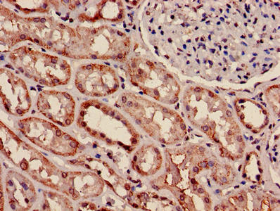
 antibody at 1:500 dilution.
Antigen Retrieval: Trilogy? (EDTA based, pH 8.0) buffer, 15min")
Non-transfected (–) and transfected (+) 293T whole cell extracts (30 μg) were separated by 5% SDS-PAGE, and the membrane was blotted with MYH9 antibody [N1], N-term (GTX101751) diluted at 1:5000. The HRP-conjugated anti-rabbit IgG antibody (GTX213110-01) was used to detect the primary antibody.
MYH9 antibody [N1], N-term
GTX101751
ApplicationsImmunoFluorescence, ImmunoPrecipitation, Western Blot, ImmunoCytoChemistry, ImmunoHistoChemistry, ImmunoHistoChemistry Paraffin
Product group Antibodies
ReactivityHuman
TargetMYH9
Overview
- SupplierGeneTex
- Product NameMYH9 antibody [N1], N-term
- Delivery Days Customer9
- Application Supplier NoteWB: 1:500-1:3000. IHC-P: 1:100-1:1000. IP: 1:100-1:500. *Optimal dilutions/concentrations should be determined by the researcher.Not tested in other applications.
- ApplicationsImmunoFluorescence, ImmunoPrecipitation, Western Blot, ImmunoCytoChemistry, ImmunoHistoChemistry, ImmunoHistoChemistry Paraffin
- CertificationResearch Use Only
- ClonalityPolyclonal
- Concentration1.31 mg/ml
- ConjugateUnconjugated
- Gene ID4627
- Target nameMYH9
- Target descriptionmyosin heavy chain 9
- Target synonymsBDPLT6, DFNA17, EPSTS, FTNS, MATINS, MHA, NMHC-II-A, NMMHC-IIA, NMMHCA, myosin-9, cellular myosin heavy chain, type A, myosin, heavy chain 9, non-muscle, non-muscle myosin heavy chain 9, non-muscle myosin heavy chain A, non-muscle myosin heavy chain IIa, non-muscle myosin heavy polypeptide 9, nonmuscle myosin IIA2, nonmuscle myosin heavy chain II-A
- HostRabbit
- IsotypeIgG
- Protein IDP35579
- Protein NameMyosin-9
- Scientific DescriptionThis gene encodes a myosin IIA heavy chain that contains an IQ domain and a myosin head-like domain. The protein is involved in several important functions, including cytokinesis, cell motility and maintenance of cell shape. Defects in MYH9 are the cause of non-syndromic sensorineural deafness autosomal dominant type 17, Epstein syndrome, Alport syndrome with macrothrombocytopenia, Sebastian syndrome, Fechtner syndrome and macrothrombocytopenia with progressive sensorineural deafness. [provided by RefSeq]
- ReactivityHuman
- Storage Instruction-20°C or -80°C,2°C to 8°C
- UNSPSC41116161
Datasheet
Related products
Product group Antibodies
Anti-MYH9 AntibodyA84609
ApplicationsWestern Blot, ELISA
ReactivityHuman, Mouse
- SizePrice
Product group Antibodies
ApplicationsWestern Blot, ELISA, ImmunoCytoChemistry
ReactivityDrosophila, Human, Mouse, Rat
TargetMYH9
- SizePrice
Product group Antibodies
Anti-MYH9 Antibody144-00173
ApplicationsWestern Blot, ImmunoHistoChemistry
ReactivityHuman, Mouse
TargetMYH9
- SizePrice
Product group Antibodies
ApplicationsWestern Blot
ReactivityHuman, Mouse, Rat
TargetMYH9
- SizePrice
Product group Antibodies
MYH9 AntibodyCSB-PA01405A0RB
ApplicationsImmunoFluorescence, ELISA, ImmunoHistoChemistry
ReactivityHuman
TargetMYH9
- SizePrice
Product group Antibodies
References

Goat anti-MYH9EB09020
ApplicationsWestern Blot, ELISA
ReactivityCanine, Human, Mouse, Rat
TargetMYH9
- SizePrice
Product group Antibodies
Myh9 Polyclonal AntibodyCAC07304
ApplicationsImmunoFluorescence, ELISA, ImmunoHistoChemistry
TargetMYH9
- SizePrice
Product group Antibodies
MYH9 antibodyGTX133377
ApplicationsWestern Blot
ReactivityHuman
TargetMYH9
- SizePrice
Product group Antibodies

MYH9 AntibodyLS-C330816
ApplicationsWestern Blot, ImmunoHistoChemistry
ReactivityHuman, Mouse
TargetMYH9
- SizePrice