

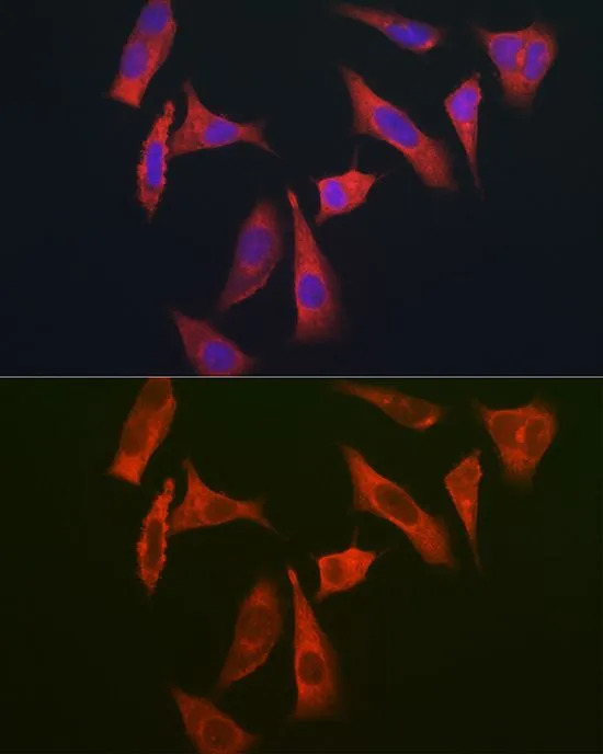
ICC/IF analysis of HeLa cells using GTX64824 MYO10 antibody. Blue : DAPI Dilution : 1:350


ICC/IF analysis of HeLa cells using GTX64824 MYO10 antibody. Blue : DAPI Dilution : 1:350
MYO10 antibody
GTX64824
ApplicationsImmunoFluorescence, Western Blot, ImmunoCytoChemistry, ImmunoHistoChemistry, ImmunoHistoChemistry Paraffin
Product group Antibodies
ReactivityHuman, Mouse
TargetMYO10
Overview
- SupplierGeneTex
- Product NameMYO10 antibody
- Delivery Days Customer9
- Application Supplier NoteWB: 1:500 - 1:2000. ICC/IF: 1:50 - 1:200. IHC-P: 1:50 - 1:200. *Optimal dilutions/concentrations should be determined by the researcher.Not tested in other applications.
- ApplicationsImmunoFluorescence, Western Blot, ImmunoCytoChemistry, ImmunoHistoChemistry, ImmunoHistoChemistry Paraffin
- CertificationResearch Use Only
- ClonalityPolyclonal
- ConjugateUnconjugated
- Gene ID4651
- Target nameMYO10
- Target descriptionmyosin X
- Target synonymsMyoX, unconventional myosin-X, unconventional myosin-10, unconventionnal myosin-X
- HostRabbit
- IsotypeIgG
- Protein IDQ9HD67
- Protein NameUnconventional myosin-X
- Scientific DescriptionThis gene encodes a member of the myosin superfamily. The protein represents an unconventional myosin; it should not be confused with the conventional non-muscle myosin-10 (MYH10). Unconventional myosins contain the basic domains of conventional myosins and are further distinguished from class members by their tail domains. This gene functions as an actin-based molecular motor and plays a role in integration of F-actin and microtubule cytoskeletons during meiosis. [provided by RefSeq, Dec 2011]
- ReactivityHuman, Mouse
- Storage Instruction-20°C or -80°C,2°C to 8°C
- UNSPSC41116161
Datasheet
Related products
Product group Antibodies
Anti-MYO10 Antibody144-12466
ApplicationsWestern Blot
ReactivityHuman, Mouse
TargetMYO10
- SizePrice

Product group Antibodies
Anti-MYO10 AntibodyAMAB91554
ApplicationsImmunoCytoChemistry
ReactivityHuman
TargetMYO10
- SizePrice
Product group Antibodies
Anti-MYO10 AntibodyA88983
ApplicationsImmunoFluorescence, Western Blot, ImmunoCytoChemistry
ReactivityHuman, Mouse
- SizePrice
Product group Antibodies

MYO10 / Myosin-X AntibodyLS-C761189
ApplicationsWestern Blot
ReactivityHuman, Mouse, Rat
TargetMYO10
- SizePrice

Product group Antibodies
MYO10 AntibodyCSB-PA884628LA01HU
ApplicationsImmunoFluorescence, ELISA, ImmunoHistoChemistry
ReactivityHuman
TargetMYO10
- SizePrice