

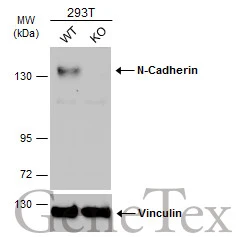
Wild-type (WT) and N-Cadherin knockout (KO) 293T cell extracts (30 μg) were separated by 5% SDS-PAGE, and the membrane was blotted with N-Cadherin antibody [N2C1], Internal (GTX112733) diluted at 1:500. The HRP-conjugated anti-rabbit IgG antibody (GTX213110-01) was used to detect the primary antibody.
![Whole cell extract (30 μg) was separated by 7.5% SDS-PAGE, and the membrane was blotted with N-Cadherin antibody [N2C1], Internal (GTX112733) diluted at 1:1000. The HRP-conjugated anti-rabbit IgG antibody (GTX213110-01) was used to detect the primary antibody.](https://www.genetex.com/upload/website/prouct_img/normal/GTX112733/GTX112733_40394_20180316_WB_w_23060500_464.webp "Whole cell extract (30 μg) was separated by 7.5% SDS-PAGE, and the membrane was blotted with N-Cadherin antibody [N2C1], Internal (GTX112733) diluted at 1:1000. The HRP-conjugated anti-rabbit IgG antibody (GTX213110-01) was used to detect the primary antibody.")
![Mouse tissue extract (50 μg) was separated by 7.5% SDS-PAGE, and the membrane was blotted with N-Cadherin antibody [N2C1], Internal (GTX112733) diluted at 1:1000.](https://www.genetex.com/upload/website/prouct_img/normal/GTX112733/GTX112733_40394_20151105_WB_M_brain_w_23060500_926.webp "Mouse tissue extract (50 μg) was separated by 7.5% SDS-PAGE, and the membrane was blotted with N-Cadherin antibody [N2C1], Internal (GTX112733) diluted at 1:1000.")
 antibody at 1:500 dilution.
Antigen Retrieval: Trilogy? (EDTA based, pH 8.0) buffer, 15min")
![Rat tissue extract (50 μg) was separated by 7.5% SDS-PAGE, and the membrane was blotted with N-Cadherin antibody [N2C1], Internal (GTX112733) diluted at 1:1000.](https://www.genetex.com/upload/website/prouct_img/normal/GTX112733/GTX112733_40394_20151105_WB_R_brain_w_23060500_519.webp "Rat tissue extract (50 μg) was separated by 7.5% SDS-PAGE, and the membrane was blotted with N-Cadherin antibody [N2C1], Internal (GTX112733) diluted at 1:1000.")
![N-Cadherin antibody [N2C1], Internal detects CDH2 protein at membrane on human hepatoma by immunohistochemical analysis. Sample: Paraffin-embedded hepatoma. N-Cadherin antibody [N2C1], Internal (GTX112733) dilution: 1:500.
Antigen Retrieval: Trilogy? (EDTA based, pH 8.0) buffer, 15min](https://www.genetex.com/upload/website/prouct_img/normal/GTX112733/GTX112733_40394_IHC_2_w_23060500_732.webp "N-Cadherin antibody [N2C1], Internal detects CDH2 protein at membrane on human hepatoma by immunohistochemical analysis. Sample: Paraffin-embedded hepatoma. N-Cadherin antibody [N2C1], Internal (GTX112733) dilution: 1:500.
Antigen Retrieval: Trilogy? (EDTA based, pH 8.0) buffer, 15min")
![N-Cadherin antibody [N2C1], Internal detects N-Cadherin protein expression by immunohistochemical analysis. Sample: Frozen sectioned E13.5 Rat brain. Green: N-Cadherin protein stained by N-Cadherin antibody [N2C1], Internal (GTX112733) diluted at 1:250. Red: beta Tubulin 3/ TUJ1, a mature neuron marker, stained by beta Tubulin 3/ TUJ1 antibody [GT11710] (GTX631836) diluted at 1:500. Blue: Fluoroshield with DAPI (GTX30920).](https://www.genetex.com/upload/website/prouct_img/normal/GTX112733/GTX112733_40394_20160921_IHC-Fr_w_23060500_134.webp "N-Cadherin antibody [N2C1], Internal detects N-Cadherin protein expression by immunohistochemical analysis. Sample: Frozen sectioned E13.5 Rat brain. Green: N-Cadherin protein stained by N-Cadherin antibody [N2C1], Internal (GTX112733) diluted at 1:250. Red: beta Tubulin 3/ TUJ1, a mature neuron marker, stained by beta Tubulin 3/ TUJ1 antibody [GT11710] (GTX631836) diluted at 1:500. Blue: Fluoroshield with DAPI (GTX30920).")
Wild-type (WT) and N-Cadherin knockout (KO) 293T cell extracts (30 μg) were separated by 5% SDS-PAGE, and the membrane was blotted with N-Cadherin antibody [N2C1], Internal (GTX112733) diluted at 1:500. The HRP-conjugated anti-rabbit IgG antibody (GTX213110-01) was used to detect the primary antibody.
N-Cadherin antibody [N2C1], Internal
GTX112733
ApplicationsImmunoFluorescence, Western Blot, ImmunoCytoChemistry, ImmunoHistoChemistry, ImmunoHistoChemistry Frozen, ImmunoHistoChemistry Paraffin
Product group Antibodies
ReactivityHuman, Mouse, Rat
TargetCDH2
Overview
- SupplierGeneTex
- Product NameN-Cadherin antibody [N2C1], Internal
- Delivery Days Customer9
- Application Supplier NoteWB: 1:500-1:3000. IHC-P: 1:100-1:1000. IHC-Fr: 1:100-1:1000. *Optimal dilutions/concentrations should be determined by the researcher.Not tested in other applications.
- ApplicationsImmunoFluorescence, Western Blot, ImmunoCytoChemistry, ImmunoHistoChemistry, ImmunoHistoChemistry Frozen, ImmunoHistoChemistry Paraffin
- CertificationResearch Use Only
- ClonalityPolyclonal
- Concentration0.53 mg/ml
- ConjugateUnconjugated
- Gene ID1000
- Target nameCDH2
- Target descriptioncadherin 2
- Target synonymsACOGS, ADHD8, ARVD14, CD325, CDHN, CDw325, NCAD, cadherin-2, N-cadherin 1, cadherin 2, type 1, N-cadherin (neuronal), calcium-dependent adhesion protein, neuronal, neural cadherin
- HostRabbit
- IsotypeIgG
- Protein IDP19022
- Protein NameCadherin-2
- Scientific DescriptionThis gene is a classical cadherin from the cadherin superfamily. The encoded protein is a calcium dependent cell-cell adhesion glycoprotein comprised of five extracellular cadherin repeats, a transmembrane region and a highly conserved cytoplasmic tail. The protein functions during gastrulation and is required for establishment of left-right asymmetry. At certain central nervous system synapses, presynaptic to postsynaptic adhesion is mediated at least in part by this gene product. [provided by RefSeq]
- ReactivityHuman, Mouse, Rat
- Storage Instruction-20°C or -80°C,2°C to 8°C
- UNSPSC41116161
Datasheet
Related products
Product group Antibodies
CDH2 AntibodyCSB-PA003379
ApplicationsImmunoFluorescence, Western Blot, ELISA, ImmunoHistoChemistry
ReactivityHuman, Mouse, Rat
TargetCDH2
- SizePrice

Product group Antibodies
Anti-N-Cadherin-2 CDH2 CD325-Antibody Picoband(r)A01577-2-CARRIER-FREE
ApplicationsWestern Blot
ReactivityHuman
TargetCDH2
- SizePrice
Product group Antibodies
Anti-CDH2 Antibody144-03045
ApplicationsImmunoPrecipitation, Western Blot
ReactivityHuman, Mouse, Rat
TargetCDH2
- SizePrice
Product group Antibodies

N-Cadherin AntibodyABX123447
ApplicationsWestern Blot, ImmunoHistoChemistry
- SizePrice

Product group Antibodies
Anti-CDH2 AntibodyAMAB91220
ApplicationsWestern Blot, ImmunoCytoChemistry, ImmunoHistoChemistry
ReactivityHuman
TargetCDH2
- SizePrice
Product group Antibodies

Anti-N-cadherin [8C11]AB01660-1.1-BT
ApplicationsFlow Cytometry, ImmunoFluorescence, Western Blot, ImmunoHistoChemistry
ReactivityHamster, Human, Rabbit
TargetCDH2
- SizePrice

Product group Antibodies
Anti-CDH2 AntibodyA98232
ApplicationsWestern Blot, ELISA
ReactivityHuman, Mouse, Rat
- SizePrice
Product group Antibodies

CDH2 / N Cadherin Antibody (FITC)LS-C488392
ApplicationsFlow Cytometry
ReactivityHuman
TargetCDH2
- SizePrice
Product group Antibodies
CDH2 Polyclonal AntibodyCAC14634
ApplicationsImmunoFluorescence, Western Blot, ELISA
ReactivityMouse
TargetCDH2
- SizePrice