

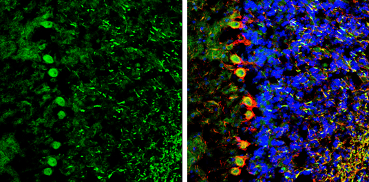
NCKAP1 antibody [C1C2], Internal detects NCKAP1 protein by immunohistochemical analysis. Sample: Frozen-sectioned mouse cerebellum. Green: NCKAP1 stained by NCKAP1 antibody [C1C2], Internal (GTX105682) diluted at 1:250. Red: NF-H, stained by NF-H antibody [GT114] (GTX634289) diluted at 1:500. Blue: Fluoroshield with DAPI (GTX30920).
![NCKAP1 antibody [C1C2], Internal detects NCKAP1 protein by Western blot analysis. A. 50 μg rat brain lysate/extract 5 % SDS-PAGE NCKAP1 antibody [C1C2], Internal (GTX105682) dilution: 1:1000](https://www.genetex.com/upload/website/prouct_img/normal/GTX105682/GTX105682_39890_WB_R_brain_w_23060120_774.webp "NCKAP1 antibody [C1C2], Internal detects NCKAP1 protein by Western blot analysis. A. 50 μg rat brain lysate/extract 5 % SDS-PAGE NCKAP1 antibody [C1C2], Internal (GTX105682) dilution: 1:1000")
 A: Mouse brain 5% SDS PAGE GTX105682 diluted at 1:1000")
![Non-transfected (–) and transfected (+) 293T whole cell extracts (30 μg) were separated by 5% SDS-PAGE, and the membrane was blotted with NCKAP1 antibody [C1C2], Internal (GTX105682) diluted at 1:5000. The HRP-conjugated anti-rabbit IgG antibody (GTX213110-01) was used to detect the primary antibody.](https://www.genetex.com/upload/website/prouct_img/normal/GTX105682/GTX105682_39890_20171215_WB_B_w_23060120_851.webp "Non-transfected (–) and transfected (+) 293T whole cell extracts (30 μg) were separated by 5% SDS-PAGE, and the membrane was blotted with NCKAP1 antibody [C1C2], Internal (GTX105682) diluted at 1:5000. The HRP-conjugated anti-rabbit IgG antibody (GTX213110-01) was used to detect the primary antibody.")
NCKAP1 antibody [C1C2], Internal detects NCKAP1 protein by immunohistochemical analysis. Sample: Frozen-sectioned mouse cerebellum. Green: NCKAP1 stained by NCKAP1 antibody [C1C2], Internal (GTX105682) diluted at 1:250. Red: NF-H, stained by NF-H antibody [GT114] (GTX634289) diluted at 1:500. Blue: Fluoroshield with DAPI (GTX30920).
NCKAP1 antibody [C1C2], Internal
GTX105682
ApplicationsWestern Blot, ImmunoHistoChemistry, ImmunoHistoChemistry Frozen
Product group Antibodies
ReactivityHuman, Mouse, Rat
TargetNCKAP1
Overview
- SupplierGeneTex
- Product NameNCKAP1 antibody [C1C2], Internal
- Delivery Days Customer9
- Application Supplier NoteWB: 1:500-1:10000. IHC-Fr: 1:100-1:1000. *Optimal dilutions/concentrations should be determined by the researcher.Not tested in other applications.
- ApplicationsWestern Blot, ImmunoHistoChemistry, ImmunoHistoChemistry Frozen
- CertificationResearch Use Only
- ClonalityPolyclonal
- Concentration0.5 mg/ml
- ConjugateUnconjugated
- Gene ID10787
- Target nameNCKAP1
- Target descriptionNCK associated protein 1
- Target synonymsHEM2, NAP1, NAP125, p125Nap1, nck-associated protein 1, membrane-associated protein HEM-2
- HostRabbit
- IsotypeIgG
- Protein IDQ9Y2A7
- Protein NameNck-associated protein 1
- Scientific DescriptionIs part of lamellipodial complex that controls Rac-dependent actin remodeling.
- ReactivityHuman, Mouse, Rat
- Storage Instruction-20°C or -80°C,2°C to 8°C
- UNSPSC12352203
Datasheet
Related products
Product group Antibodies
Anti-NCKAP1 Antibody144-12229
ApplicationsWestern Blot
ReactivityHuman, Mouse, Rat
TargetNCKAP1
- SizePrice
Product group Antibodies

NCKAP1 / NAP125 AntibodyLS-C747386
ApplicationsWestern Blot
ReactivityHuman, Mouse, Rat
TargetNCKAP1
- SizePrice
Product group Antibodies
Anti-NCKAP1 AntibodyA326266
ApplicationsFlow Cytometry, ImmunoFluorescence, ELISA
ReactivityHuman
- SizePrice
Product group Antibodies

Goat anti-NCKAP1 AntibodyEB11695
ApplicationsFlow Cytometry, ImmunoFluorescence, ELISA
ReactivityBovine, Canine, Human, Mouse, Porcine, Rat
TargetNCKAP1
- SizePrice
Product group Antibodies
NCKAP1 AntibodyCSB-PA295755
ApplicationsELISA, ImmunoHistoChemistry
ReactivityHuman, Mouse, Rat
TargetNCKAP1
- SizePrice

Product group Antibodies
Anti-NCKAP1 AntibodyHPA020449
ApplicationsImmunoCytoChemistry, ImmunoHistoChemistry
ReactivityHuman
TargetNCKAP1
- SizePrice
Product group Antibodies
NCKAP1 antibody [N1N2], N-termGTX115481
ApplicationsWestern Blot, ImmunoHistoChemistry, ImmunoHistoChemistry Frozen, ImmunoHistoChemistry Paraffin
ReactivityHuman, Mouse, Rat
TargetNCKAP1
- SizePrice

Product group Antibodies
Anti-NCKAP1 Antibody Picoband(r)A09238-1-CARRIER-FREE
ApplicationsWestern Blot
ReactivityHuman, Mouse, Rat
TargetNCKAP1
- SizePrice