

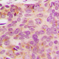
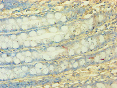
IHC-P analysis of formalin fixed human breast cancer tissue section using GTX56108 NDUFA3 antibody. Antigen retrieval : Heat mediated antigen retrieval with sodium citrate buffer (pH 6.0)
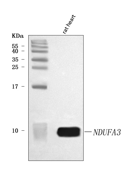
, Raw264.7 (B) whole cell lysates using GTX56108 NDUFA3 antibody.")
IHC-P analysis of formalin fixed human breast cancer tissue section using GTX56108 NDUFA3 antibody. Antigen retrieval : Heat mediated antigen retrieval with sodium citrate buffer (pH 6.0)
NDUFA3 antibody
GTX56108
ApplicationsWestern Blot, ImmunoHistoChemistry, ImmunoHistoChemistry Paraffin
Product group Antibodies
ReactivityHuman, Mouse
TargetNDUFA3
Overview
- SupplierGeneTex
- Product NameNDUFA3 antibody
- Delivery Days Customer9
- Application Supplier NoteWB: 1:500 - 1:1000. IHC-P: 1:100 - 1:200. *Optimal dilutions/concentrations should be determined by the researcher.Not tested in other applications.
- ApplicationsWestern Blot, ImmunoHistoChemistry, ImmunoHistoChemistry Paraffin
- CertificationResearch Use Only
- ClonalityPolyclonal
- ConjugateUnconjugated
- Gene ID4696
- Target nameNDUFA3
- Target descriptionNADH:ubiquinone oxidoreductase subunit A3
- Target synonymsB9, CI-B9, NADH dehydrogenase [ubiquinone] 1 alpha subcomplex subunit 3, NADH dehydrogenase (ubiquinone) 1 alpha subcomplex, 3, 9kDa, NADH-ubiquinone oxidoreductase B9 subunit, complex I B9 subunit, complex I-B9
- HostRabbit
- IsotypeIgG
- Protein IDO95167
- Protein NameNADH dehydrogenase [ubiquinone] 1 alpha subcomplex subunit 3
- ReactivityHuman, Mouse
- Storage Instruction-20°C or -80°C,2°C to 8°C
- UNSPSC12352203
Datasheet
Related products
Product group Antibodies
Anti-NDUFA3 Antibody Picoband(r)A11694-1-CARRIER-FREE
ApplicationsWestern Blot, ELISA
ReactivityHuman, Rat
TargetNDUFA3
- SizePrice

Product group Antibodies
Anti-NDUFA3 AntibodyA98629
ApplicationsELISA, ImmunoHistoChemistry
ReactivityHuman, Mouse
- SizePrice
Product group Antibodies
NDUFA3 AntibodyCSB-PA015624EA01HU
ApplicationsImmunoFluorescence, ELISA, ImmunoHistoChemistry
ReactivityHuman
TargetNDUFA3
- SizePrice

Product group Antibodies
Anti-NDUFA3 AntibodyHPA046976
ApplicationsImmunoHistoChemistry
ReactivityHuman
TargetNDUFA3
- SizePrice
Product group Antibodies

NDUFA3 / B9 AntibodyLS-C285566
ApplicationsWestern Blot, ELISA
ReactivityHuman
TargetNDUFA3
- SizePrice