

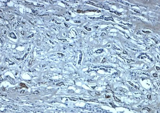
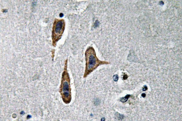
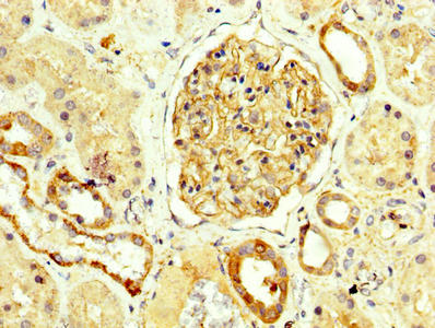
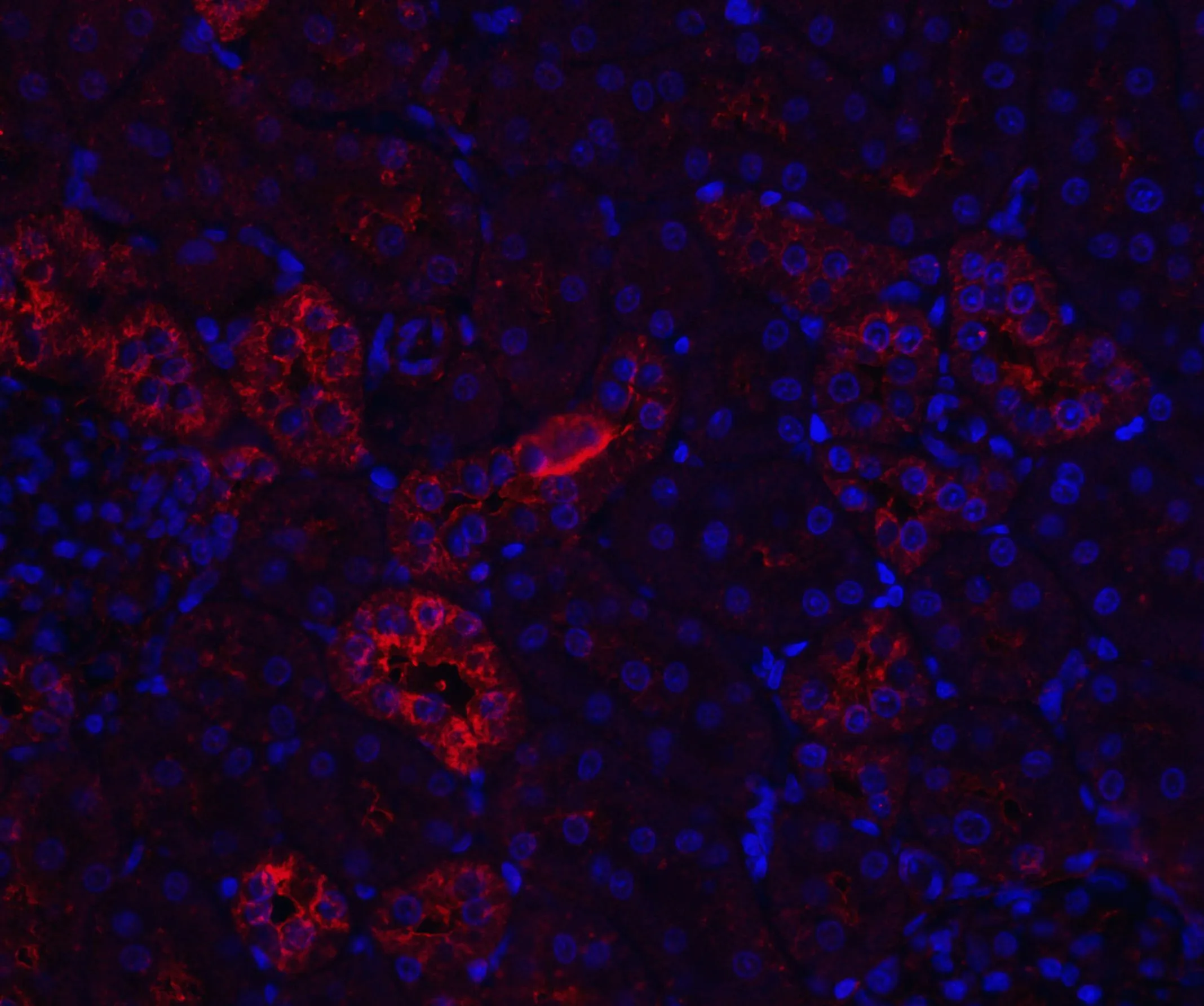
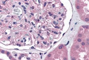
IHC-P analysis of human kidney tissue using GTX31936 Nephrin antibody. Working concentration : 5 μg/ml
 the absence and (B) the presence of blocking peptide using GTX31936 Nephrin antibody. Working concentration : 1 μg/ml")

IHC-P analysis of human kidney tissue using GTX31936 Nephrin antibody. Working concentration : 5 μg/ml
Nephrin antibody
GTX31936
ApplicationsWestern Blot, ELISA, ImmunoHistoChemistry, ImmunoHistoChemistry Paraffin
Product group Antibodies
ReactivityHuman
TargetNPHS1
Overview
- SupplierGeneTex
- Product NameNephrin antibody
- Delivery Days Customer9
- Application Supplier NoteWB: 1 - 2 microg/mL. IHC-P: 5 microg/mL. *Optimal dilutions/concentrations should be determined by the researcher.Not tested in other applications.
- ApplicationsWestern Blot, ELISA, ImmunoHistoChemistry, ImmunoHistoChemistry Paraffin
- CertificationResearch Use Only
- ClonalityPolyclonal
- Concentration1 mg/ml
- ConjugateUnconjugated
- Gene ID4868
- Target nameNPHS1
- Target descriptionNPHS1 adhesion molecule, nephrin
- Target synonymsCNF, NPHN, nephrin, nephrin, NPHS1, nephrin, nephrosis 1, congenital, Finnish type (nephrin), renal glomerulus-specific cell adhesion receptor
- HostRabbit
- IsotypeIgG
- Protein IDO60500
- Protein NameNephrin
- Scientific DescriptionThis gene encodes a member of the immunoglobulin family of cell adhesion molecules that functions in the glomerular filtration barrier in the kidney. The gene is primarily expressed in renal tissues, and the protein is a type-1 transmembrane protein found at the slit diaphragm of glomerular podocytes. The slit diaphragm is thought to function as an ultrafilter to exclude albumin and other plasma macromolecules in the formation of urine. Mutations in this gene result in Finnish-type congenital nephrosis 1, characterized by severe proteinuria and loss of the slit diaphragm and foot processes.[provided by RefSeq, Oct 2009]
- ReactivityHuman
- Storage Instruction-20°C or -80°C,2°C to 8°C
- UNSPSC41116161
References
- Dendrobium officinale polysaccharide decreases podocyte injury in diabetic nephropathy by regulating IRS-1/AKT signal and promoting mitophagy.Read this paper
Datasheet
Related products
Product group Antibodies
Anti-Nephrin AntibodyA96354
ApplicationsELISA, ImmunoHistoChemistry
ReactivityHuman, Mouse, Rat
- SizePrice
Product group Antibodies
Anti-NPHS1 Antibody144-65639
ApplicationsImmunoFluorescence, Western Blot, ImmunoHistoChemistry
ReactivityHuman, Mouse, Rat
TargetNPHS1
- SizePrice

Product group Antibodies
Anti-Nephrin/NPHS1 Antibody Picoband(r)A01991-CARRIER-FREE
ApplicationsWestern Blot, ImmunoHistoChemistry
ReactivityHuman, Mouse, Rat
TargetNPHS1
- SizePrice

Product group Antibodies
References
Nephrin Polyclonal AntibodyBS-10233R
ApplicationsFlow Cytometry, ImmunoFluorescence, Western Blot, ImmunoHistoChemistry, ImmunoHistoChemistry Frozen, ImmunoHistoChemistry Paraffin
ReactivityHuman, Rabbit
TargetNPHS1
- SizePrice
Product group Antibodies
Nphs1 Polyclonal AntibodyCAC08678
ApplicationsImmunoFluorescence, Western Blot, ELISA, ImmunoHistoChemistry
TargetNPHS1
- SizePrice
Product group Antibodies
NPHS1 AntibodyCSB-PA015988LA01HU
ApplicationsImmunoFluorescence, Western Blot, ELISA, ImmunoHistoChemistry
ReactivityHuman
TargetNPHS1
- SizePrice
Product group Antibodies
References
Nephrin antibodyGTX31654
ApplicationsWestern Blot, ELISA, ImmunoHistoChemistry, ImmunoHistoChemistry Paraffin
ReactivityHuman, Mouse, Rat
TargetNPHS1
- SizePrice
Product group Antibodies
Nephrin antibodyGTX31799
ApplicationsELISA, ImmunoHistoChemistry, ImmunoHistoChemistry Paraffin
ReactivityHuman
TargetNPHS1
- SizePrice