

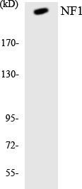
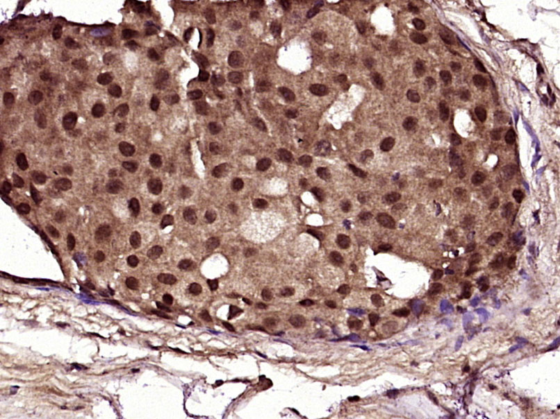
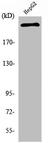
NF1 Rabbit pAb, FITC conjugated
ORB103091
ApplicationsFlow Cytometry, ImmunoFluorescence, ImmunoCytoChemistry
Product group Antibodies
ReactivityCanine, Equine, Human, Mouse, Rabbit, Rat
Overview
- SupplierBiorbyt
- Product NameNF1 Rabbit pAb, FITC conjugated
- Delivery Days Customer16
- ApplicationsFlow Cytometry, ImmunoFluorescence, ImmunoCytoChemistry
- Applications SupplierFlow-Cyt=1μg/Test, IF=1:100-500, ICC/IF=1:100 FC, ICC, IF
- CertificationResearch Use Only
- ClonalityPolyclonal
- Concentration1 mg/ml
- ConjugateFITC
- HostRabbit
- IsotypeIgG
- Scientific DescriptionNF1 Rabbit pAb, FITC conjugated
- ReactivityCanine, Equine, Human, Mouse, Rabbit, Rat
- Storage Instruction-20°C
- UNSPSC12352203
Related products
Product group Antibodies
Anti-NF1 AntibodyA98470
ApplicationsWestern Blot, ELISA
ReactivityHuman, Mouse, Rat
- SizePrice

Product group Antibodies
Anti-NF1 AntibodyAMAB91741
ApplicationsImmunoCytoChemistry
ReactivityHuman
TargetNF1
- SizePrice
Product group Antibodies

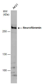
Neurofibromin / NF1 AntibodyLS-C832719
ApplicationsELISA, ImmunoHistoChemistry
ReactivityHuman, Mouse, Rat
TargetNF1
- SizePrice

Product group Antibodies
Anti-Neurofibromin/NF1 Antibody Picoband(r)A00043-1-CARRIER-FREE
ApplicationsFlow Cytometry, ImmunoFluorescence, Western Blot, ELISA, ImmunoCytoChemistry, ImmunoHistoChemistry
ReactivityHuman
TargetNF1
- SizePrice
Product group Antibodies
NF1 Polyclonal AntibodyBS-4140R
ApplicationsImmunoFluorescence, ELISA, ImmunoCytoChemistry, ImmunoHistoChemistry, ImmunoHistoChemistry Frozen, ImmunoHistoChemistry Paraffin
ReactivityCanine, Equine, Human, Mouse, Rabbit, Rat
TargetNF1
- SizePrice
Product group Antibodies
NF1 AntibodyCSB-PA003413
ApplicationsImmunoFluorescence, Western Blot, ELISA
ReactivityHuman, Mouse, Rat
TargetNF1
- SizePrice
Product group Antibodies
Neurofibromin antibodyGTX132099
ApplicationsWestern Blot
ReactivityHuman
TargetNF1
- SizePrice