

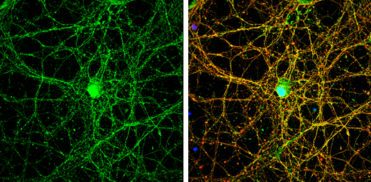
NMDAR2B antibody [C2C3], C-term detects NMDAR2B protein by immunofluorescent analysis. Sample: DIV14 rat E18 primary cortical neurons were fixed in 4% paraformaldehyde at RT for 15 min. Green: NMDAR2B protein stained by NMDAR2B antibody [C2C3], C-term (GTX109713) diluted at 1:500. Red: beta Tubulin 3/ Tuj1, stained by beta Tubulin 3/ Tuj1 antibody [GT1338] (GTX631831) diluted at 1:500. Blue: Fluoroshield with DAPI (GTX30920).
 A: Hep G2 (GTX27900) 5% SDS PAGE GTX109713 diluted at 1:1000")
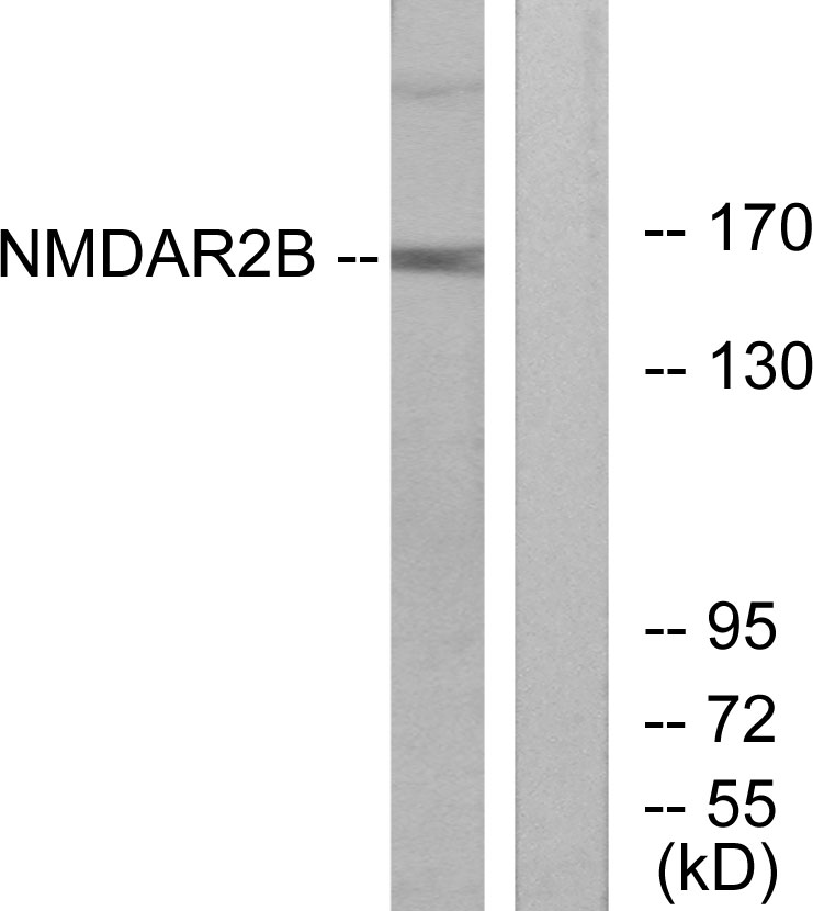
![Various tissue extracts (50 μg) were separated by 5% SDS-PAGE, and the membrane was blotted with NMDAR2B antibody [C2C3], C-term (GTX109713) diluted at 1:3000. The HRP-conjugated anti-rabbit IgG antibody (GTX213110-01) was used to detect the primary antibody.](https://www.genetex.com/upload/website/prouct_img/normal/GTX109713/GTX109713_40023_20170706_WB_M_R_w_23060500_265.webp "Various tissue extracts (50 μg) were separated by 5% SDS-PAGE, and the membrane was blotted with NMDAR2B antibody [C2C3], C-term (GTX109713) diluted at 1:3000. The HRP-conjugated anti-rabbit IgG antibody (GTX213110-01) was used to detect the primary antibody.")
NMDAR2B antibody [C2C3], C-term detects NMDAR2B protein by immunofluorescent analysis. Sample: DIV14 rat E18 primary cortical neurons were fixed in 4% paraformaldehyde at RT for 15 min. Green: NMDAR2B protein stained by NMDAR2B antibody [C2C3], C-term (GTX109713) diluted at 1:500. Red: beta Tubulin 3/ Tuj1, stained by beta Tubulin 3/ Tuj1 antibody [GT1338] (GTX631831) diluted at 1:500. Blue: Fluoroshield with DAPI (GTX30920).
NMDAR2B antibody [C2C3], C-term
GTX109713
ApplicationsImmunoFluorescence, Western Blot, ImmunoCytoChemistry
Product group Antibodies
ReactivityHuman, Mouse, Rat
TargetGRIN2B
Overview
- SupplierGeneTex
- Product NameNMDAR2B antibody [C2C3], C-term
- Delivery Days Customer9
- Application Supplier NoteWB: 1:500-1:3000. ICC/IF: 1:100-1:1000. *Optimal dilutions/concentrations should be determined by the researcher.Not tested in other applications.
- ApplicationsImmunoFluorescence, Western Blot, ImmunoCytoChemistry
- CertificationResearch Use Only
- ClonalityPolyclonal
- Concentration0.79 mg/ml
- ConjugateUnconjugated
- Gene ID2904
- Target nameGRIN2B
- Target descriptionglutamate ionotropic receptor NMDA type subunit 2B
- Target synonymsDEE27, EIEE27, GluN2B, MRD6, NMDAR2B, NR2B, NR3, hNR3, glutamate receptor ionotropic, NMDA 2B, GluN2B(alt_5'UTR), N-methyl D-aspartate receptor subtype 2B, N-methyl-D-aspartate receptor subunit 3, glutamate [NMDA] receptor subunit epsilon-2, glutamate receptor subunit epsilon-2, glutamate receptor, ionotropic, N-methyl D-aspartate 2B
- HostRabbit
- IsotypeIgG
- Protein IDQ13224
- Protein NameGlutamate receptor ionotropic, NMDA 2B
- Scientific DescriptionN-methyl-D-aspartate (NMDA) receptors are a class of ionotropic glutamate receptors. NMDA receptor channel has been shown to be involved in long-term potentiation, an activity-dependent increase in the efficiency of synaptic transmission thought to underlie certain kinds of memory and learning. NMDA receptor channels are heteromers composed of three different subunits: NR1 (GRIN1), NR2 (GRIN2A, GRIN2B, GRIN2C, or GRIN2D) and NR3 (GRIN3A or GRIN3B). The NR2 subunit acts as the agonist binding site for glutamate. This receptor is the predominant excitatory neurotransmitter receptor in the mammalian brain. [provided by RefSeq]
- ReactivityHuman, Mouse, Rat
- Storage Instruction-20°C or -80°C,2°C to 8°C
- UNSPSC41116161
Datasheet
Related products
Product group Antibodies
Anti-NMDAR2B AntibodyA96564
ApplicationsWestern Blot, ELISA
ReactivityHuman, Mouse, Rat
- SizePrice
Product group Antibodies
Anti-GRIN2B Antibody144-65641
ApplicationsImmunoFluorescence, Western Blot
ReactivityHuman, Mouse, Rat
TargetGRIN2B
- SizePrice
Product group Antibodies
ApplicationsImmunoFluorescence, Western Blot, ELISA, ImmunoCytoChemistry, ImmunoHistoChemistry, ImmunoHistoChemistry Frozen, ImmunoHistoChemistry Paraffin
ReactivityBovine, Canine, Chicken, Equine, Human, Mouse, Rat
TargetGRIN2B
- SizePrice
Product group Antibodies
Phospho-GRIN2B (S1303) AntibodyCSB-PA008978
ApplicationsWestern Blot, ELISA
ReactivityHuman, Mouse, Rat
TargetGRIN2B
- SizePrice
Product group Antibodies

Goat anti-NMDAR2B / GRIN2BEB07294
ApplicationsImmunoFluorescence, Western Blot, ELISA
ReactivityCanine, Human, Mouse, Rat
TargetGRIN2B
- SizePrice
Product group Antibodies
ApplicationsImmunoPrecipitation, Western Blot, ImmunoCytoChemistry, ImmunoHistoChemistry
ReactivityMouse, Porcine, Rat
TargetGRIN2B
- SizePrice
Product group Antibodies

GRIN2B / NMDAR2B / NR2B AntibodyLS-C405753
ApplicationsWestern Blot, ELISA
ReactivityHuman, Mouse, Rat
TargetGRIN2B
- SizePrice
![IHC-P analysis of human breast tissue using GTX80691 NMDAR2B antibody [NR2B]. Right : Primary antibody Left : Negative control without primary antibody Antigen retrieval : 10mM sodium citrate (pH 6.0), microwaved for 8-15 min Dilution : 1:20](https://www.genetex.com/upload/website/prouct_img/normal/GTX80691/GTX80691_1408_IHC-P_w_23061322_716.webp)
Product group Antibodies
NMDAR2B antibody [NR2B]GTX80691
ApplicationsImmunoFluorescence, Western Blot, ImmunoCytoChemistry, ImmunoHistoChemistry, ImmunoHistoChemistry Paraffin
ReactivityHuman, Mouse, Rat
TargetGRIN2B
- SizePrice
Product group Antibodies
NMDAR2B antibody, InternalGTX89310
ApplicationsWestern Blot
ReactivityHuman, Rat
TargetGRIN2B
- SizePrice

Product group Antibodies
Anti-GRIN2B AntibodyHPA069762
ApplicationsImmunoHistoChemistry
ReactivityHuman
TargetGRIN2B
- SizePrice