

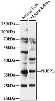
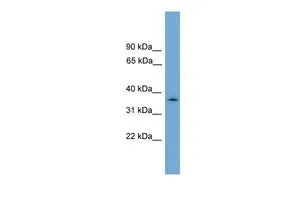
NUBP1 Polyclonal Antibody
BS-7817R
ApplicationsImmunoFluorescence, Western Blot, ELISA, ImmunoCytoChemistry, ImmunoHistoChemistry, ImmunoHistoChemistry Frozen, ImmunoHistoChemistry Paraffin
Product group Antibodies
ReactivityBovine, Canine, Chicken, Equine, Human, Mouse, Porcine, Rat, Sheep
TargetNUBP1
Overview
- SupplierBioss
- Product NameNUBP1 Polyclonal Antibody
- Delivery Days Customer16
- ApplicationsImmunoFluorescence, Western Blot, ELISA, ImmunoCytoChemistry, ImmunoHistoChemistry, ImmunoHistoChemistry Frozen, ImmunoHistoChemistry Paraffin
- Applications SupplierWB(1:300-5000), ELISA(1:500-1000), IHC-P(1:200-400), IHC-F(1:100-500), IF(IHC-P)(1:50-200), IF(IHC-F)(1:50-200), IF(ICC)(1:50-200)
- CertificationResearch Use Only
- ClonalityPolyclonal
- Concentration1 ug/ul
- ConjugateUnconjugated
- Gene ID4682
- Target nameNUBP1
- Target descriptionNUBP iron-sulfur cluster assembly factor 1, cytosolic
- Target synonymsCIAO5, NBP, NBP1, NBP35, cytosolic Fe-S cluster assembly factor NUBP1, NBP 1, nucleotide binding protein (e.coli MinD like), nucleotide binding protein 1 (E.coli MinD like), nucleotide binding protein 1 (MinD homolog, E. coli), nucleotide-binding protein 1
- HostRabbit
- IsotypeIgG
- ReactivityBovine, Canine, Chicken, Equine, Human, Mouse, Porcine, Rat, Sheep
- Storage Instruction-20°C
- UNSPSC12352203
Datasheet
Related products

Product group Antibodies
Anti-NUBP1 Antibody Picoband(r)A08538-1-CARRIER-FREE
ApplicationsFlow Cytometry, ImmunoFluorescence, Western Blot, ELISA, ImmunoCytoChemistry
ReactivityHuman
TargetNUBP1
- SizePrice
Product group Antibodies
Anti-NUBP1 Antibody144-64348
ApplicationsWestern Blot, ImmunoHistoChemistry
ReactivityHuman, Mouse, Rat
TargetNUBP1
- SizePrice
Product group Antibodies
NUBP1 AntibodyCSB-PA016142XA01HU
ApplicationsWestern Blot, ELISA
ReactivityHuman
TargetNUBP1
- SizePrice

Product group Antibodies
Anti-NUBP1 AntibodyHPA041656
ApplicationsWestern Blot, ImmunoHistoChemistry
ReactivityHuman
TargetNUBP1
- SizePrice
Product group Antibodies

NBP / NUBP1 AntibodyLS-C833748
ApplicationsImmunoHistoChemistry
ReactivityHuman, Mouse, Rat
TargetNUBP1
- SizePrice
Product group Antibodies
NUBP1 antibody, N-termGTX45063
ApplicationsWestern Blot
ReactivityHuman
TargetNUBP1
- SizePrice